Global collaboration is not a thing of the future. It is already here. The development of international friendships and collaborations will help to reduce the burden of preventable glaucoma blindness. In this edition of “International Perspectives,” Ganesh Venkataraman, MBBS, MS, DNB, shares his experiences observing top-tier ophthalmology programs, something hardly any of us has had the privilege of doing since applying for either residency or fellowship. Top-tier academic, clinical, and basic research programs in the developing world often place a strong emphasis on community service. Global collaborations offer the opportunity to merge the best possible elements of ophthalmic training and hold the possibility of innovating glaucoma treatment.
—Alan L. Robin, MD, section editor
I would like to share my experiences observing academic medicine at various institutions in the United States. Thanks to both my parent organization, the Aravind Eye Care System in Tamil Nadu, India, and to the Seva Foundation, which in part financially supported my attendance at the 2014 Annual Meeting of the American Academy of Ophthalmology through the Host an Ophthalmologist Program, I had the opportunity to visit various US academic eye care centers. I chose four diverse institutions, each of which has glaucoma specialists who have collaborated with Aravind. I observed the daily functioning of the Glaucoma Services at Johns Hopkins University, the University of Michigan, the University of Iowa, and the University of Miami. I was treated graciously and supported by the faculty members at each of these institutions.
FIRST STOP: MARYLAND
Pradeep Ramulu, MD, MPH, was my host at the Wilmer Eye Institute at Johns Hopkins University. While there, I observed both the clinic and OR. The faculty is assisted by residents and fellows, and I was exposed to the intense supervision and training the students receive.
On Thursday mornings, most of the Wilmer faculty members participate in grand rounds. First- and second-year residents are tasked with presenting their most fascinating cases. Afterward, a 3-hour didactic session combines a lecture, small group discussion, and hands-on learning.
I was especially impressed with the weekly Professor Rounds meeting, where residents and Chairman Peter J. McDonnell, MD, come together. One resident per session presents on a topic. The chief resident moderates the discussion, and Dr. McDonnell shares his relevant experience. I thought this process was an excellent opportunity for residents to interact with Dr. McDonnell, apart from their brief clinical rotation with him.
Each subspecialty division also holds journal clubs and conferences throughout the year at which residents are invited to attend and give presentations. Based on my observations, these are informal, small gatherings, sometimes even held at a attendings’ homes.
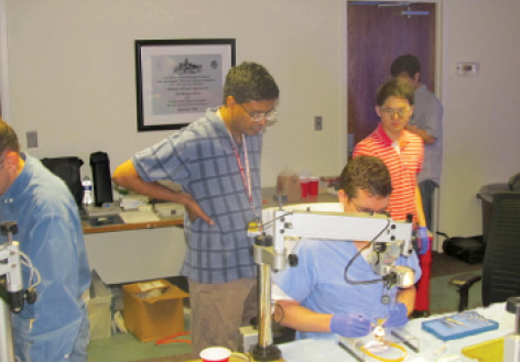
Figure 1. Dr. Ramulu assists residents in the tube laboratory.
Dr. Ramulu conducted wet lab instruction for first- and second-year residents to practice glaucoma drainage device surgery (Figure 1). A lecture preceded the wet lab portion. At Aravind, we have experience with implanting the Aurolab Aqueous Drainage Implant (AADI), which is similar to the Baerveldt glaucoma implant (Abbott Medical Optics). A tube wet lab would be a great idea for Aravind’s fellows. I learned firsthand at Wilmer the importance of glaucoma drainage device wet labs and how to conduct one.
SECOND STOP: MICHIGAN
Residents and fellows at the Kellogg Eye Center at the University of Michigan are fortunate to work with a large faculty well known for international collaboration with health care providers in less privileged parts of the world such as Ethiopia, Ghana, India, and South America. International collaboration is nurtured through the University’s Global REACH (Research, Education, and Collaboration in Health) Department, which encourages students to participate in health programs with collaborators around the world.
This year, at Kellogg’s annual International Night, Dr. Sanduk Ruit, a founder of the Tilganga Eye Hospital, Magsaysay Award winner, and cofounder of the Himalayan Cataract Project, presented on eye care projects and services in Nepal (Figure 2). Students presented posters and gave lectures at the event.
Bennie Jeng, MD, MS, chairman of the University of Maryland School of Medicine’s Department of Ophthalmology and Visual Sciences, presented on recent advances in the management of corneal diseases at a weekly grand rounds meeting.
Residents and fellows assist faculty in advanced surgical procedures, including seton surgery, canaloplasty, and phacoemulsification. They also perform these procedures under close supervision. After clinic, the faculty and residents come together to discuss the day’s interesting clinical cases.
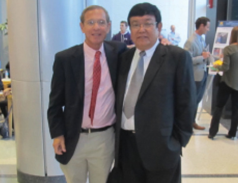
Figure 2. Dr. Robin with Dr. Ruit.
An intriguing feature at Kellogg is the Center of Ophthalmic Photography, which provides in-house training for technicians learning imaging. The training process starts at the front desk. The trainee slowly learns the nuances of external digital photography and progresses to imaging the internal structures of the eye using sophisticated instruments such as the fundus camera and optical coherence tomography. These are lessons that will be important for our collaborations on screening and telemedicine.
THIRD STOP: IOWA
With excellent facilities for imaging technology and an easily accessible as well as large genetics laboratory in the same building, the Univeristy of Iowa’s strength is developing clinician scientists. Unlike at the other schools I visited, grand rounds are held daily. Faculty lectures take place once a month, and residents attend a lecture and discussion on clinical genetics twice a week. Fellows have an opportunity to work with several faculty members, and they rotate every 3 to 4 months among professors.
The residents and fellows assist the faculty with surgical procedures such as iridocyclectomies, setons, tube implants, and angle surgeries. In addition to routine clinical discussions, the Glaucoma Service features the discussion of a daily topic. Subjects range from aqueous humour dynamics to the management of intractable glaucoma. Residents can choose to attend topic discussions according to their interests.
From the vast collection of recorded videos and photographs meticulously catalogued by Lee Alward, MD, the residents and fellows have access to numerous clinical presentations of the same condition. Genetic research and organizational skills were the key take-away messages.
FOURTH STOP: FLORIDA
My last stop was at the Bascom Palmer Eye Institute in Miami. Ever since Aurolab (India) developed the AADI, there has been a mutual exchange of ideas and faculty visits between Bascom Palmer and Aravind. Paul Palmberg, MD, PhD, visited us in 2011 and personally taught me how to implant the AADI. Since his retirement, Dr. Palmberg spends his time helping the residents with their clinics. He also assists residents in the OR. The involvement of such an experienced and eminent glaucoma surgeon is a benefit that very few centers’ residency programs can boast.
Weekly grand rounds, moderated by Chairman Eduardo Alfonso, MD, include three presentations. Interestingly, all presenters are required to submit an abstract to a peer-reviewed journal.
Bascom Palmer welcomes visiting ophthalmologists from South America. The Annual Inter-American Course in Clinical Ophthalmology, conducted by the university, invites participants from Latin American countries to interact with their US counterpart. I was fortunate to attend the glaucoma session, where I heard new ideas about shunts and procedures such as gonioscopy-assisted transluminal trabeculoplasty.
CONCLUSION
Many of my colleagues are keen to know which center I thought was the best. All had many strengths and faced challenges for smooth functioning. Each center managed to generate innovative ideas for satisfying of patients, whether it be a “pod” layout of the consultation suite at Bascom Palmer or having the genetics research laboratory attached to the main hospital wing at the University of Iowa. The faculty at every center worked in close collaboration with each other toward the primary goal of enhancing patients’ care in clinics as well as through research. Student and fellow teaching methods were very hands-on, with one-to-one teaching in the clinics and in the OR. With effective faculty-to-student ratios, great audiovisual aids, and appropriate use of technology (simple things like a teaching microscope in the slit lamp), the resident and fellow teaching programs at these academic centers are the most excellent and obviously much sought after. A visit to any one of the centers would broaden the horizons of residents or fellows from less privileged regions. n
Section Editor Alan L. Robin, MD, is an associate professor of ophthalmology at the Wilmer Eye Institute and an associate professor of international health at the Bloomberg School of Public Health, both at Johns Hopkins University in Baltimore. He is also a professor at the University of Maryland and an adjunct professor at the University of Michigan. Dr. Robin may be reached at (410) 377-2422; arobin@glaucomaexpert.com.
Ganesh Venkataraman, MBBS, MS, DNB, is chief consultant, glaucoma, at Aravind Eye Hospital in Coimbatore, India. He acknowledged no financial interest in the products or companies mentioned herein. Dr. Venkataraman may be reached at ganeshvr75@gmail.com.