The year 2020 marked the 200th anniversary of the founding of the New York Eye and Ear Infirmary of Mount Sinai (NYEE)—a significant milestone made more memorable by the concurrent global pandemic. Amid great disruption in New York City—a US epicenter of the COVID-19 outbreak—our team at NYEE was able to bring to the institution (and to the United States in general) a first-in-class robotic system for ophthalmic surgery (Figure 1).
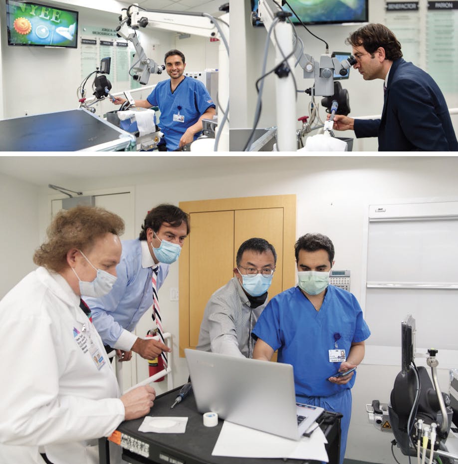
Figure 1. Members of the NYEE robotics team work to implement and adapt the Preceyes Surgical System.
The Preceyes Surgical System (Preceyes; Figure 2) is designed to support surgeons in optimizing ophthalmic surgery and assist in the development of novel high-precision treatments. The platform received the CE Mark in 2019 for external membrane peeling and has been used successfully for vitreoretinal surgery in approximately 50 eyes to date (data on file, Preceyes).

Figure 2. The Preceyes Surgical System scales movements and filters tremors of the surgeon, thereby increasing precision and stability.
This article explores the potential of this breakthrough technology for surgical glaucoma care and the increased precision it could bring.
MINIMIZING MICROSURGERY
Skilled ophthalmic surgeons routinely achieve satisfactory outcomes with manual surgery. However, manual vitreoretinal surgery has been reported to have a root mean square amplitude of tremor of 182 µm and a peak-to-peak vector magnitude of 100 µm.1 Upon considering the size of various ophthalmic structures—120 µm for macular holes,2 100 to 150 µm for retinal veins,3 and 100 to 150 µm for Schlemm canal4—it becomes clear that ophthalmologists operate at the limit of anatomic measurements.
MIGS in particular has enabled ophthalmic surgery to reach new levels of precision with small hardware. MIGS stents continue to decrease in size, and MIGS surgeons achieve ab interno access to very small structures such as the trabecular meshwork (approximately 150 µm).
Precision matters with MIGS. Figure 3 shows a postoperative image of one of my patients whose IOP seemed well controlled after implantation of a MIGS stent. However, gonioscopic examination revealed that the stent was located below the trabecular meshwork. Although the device was successfully implanted, it was not in the appropriate anatomic position to be functional.

Within the FDA Manufacturer and User Facility Device Experience database of adverse events,5 the rate of malposition with some MIGS stents is reported to be around 30%. It is important to consider how this relates to safety and efficacy and whether more precise implantation could make a significant difference—this is where robotics comes into play. Precision tremor stabilization decreases from between 120 µm and 150 µm manually to less than 5 µm with robotics.6 Additional capabilities such as smart instrumentation, pressure sensing, and integration of data and analytics could further increase the level of precision achieved with robotics.
BRINGING ROBOTICS TO GLAUCOMA SURGERY
The Preceyes Surgical System consists of a manipulator to hold a surgical instrument and a joystick that the surgeon uses to control the movements of the instrument. In practice, the manipulator copies the movements of the surgeon, leaving the surgeon in full control of the instrument and the surgery. The device scales movements and filters tremors of the surgeon, thereby increasing precision and stability.
Our team at the NYEE Robotics Program sought to apply the Preceyes Surgical System to glaucoma surgery—goniotomy and iStent (Glaukos) implantation specifically—but some modifications were required. We used a 3D printer to create an adapter for coupling the iStent injector with the robot. We then worked to adapt the angle of approach needed for adequate visualization and determine the parameters for operating a high-precision device in the angle using a joystick.
Ultimately, we were able to successfully perform high-precision goniotomy and iStent implantation in a synthetic eye. The robot was used for anterior chamber entry, engagement of the trabecular meshwork, implantation and deployment of the iStent, and high-precision goniotomy excision. Our experience operating the Preceyes device validated the reported lack of tremor and the high level of precision possible with its use.
CONCLUSION
Great collaboration was required to bring the Preceyes Surgical System to the United States and modify it for use in glaucoma surgery. We look forward to continuing to investigate where new levels of precision can take ophthalmic surgery in the future and seeing how these advances can improve patient outcomes. Further, with the Preceyes connected to the EyeSi surgical simulator, significant opportunities for training the next generation of microsurgeons abound.
1. Singhy SPN, Riviere C. Physiological tremor amplitude during retinal microsurgery. Paper presented at: Annual International Conference of the IEEE Engineering in Medicine and Biology Society; 2002.
2. Shin JY, Chu YK, Hong YT, Kwon OW, Byeon SH. Determination of macular hole size in relation to individual variabilities of fovea morphology. Eye (Lond). 2015;29(8):1051-1059.
3. Goldenberg D. Shahar J, Loewenstein A, Goldstein M. Diameters of retinal blood vessels in a healthy cohort as measured by spectral domain optical coherence tomography. Retina. 2013;33(9):1888-1894.
4. Irshad FA, Mayfield MS, Zurakowski D, Ayyala RS. Variation in Schlemm’s canal diameter and location by ultrasound biomicroscopy. Ophthalmology. 2010;117(5):916-920.
5. Duong AT, Yuan M, Koenig LR, Rodriguez GH, Van Tassel SH. Adverse events associated with microinvasive glaucoma surgery reported to the Food and Drug Administration. Ophthalmol Glaucoma. 2021;4(4):433-435.
6. de Smet MD, de Jonge N, Iannetta D, et al. Human/robotic interaction: vision limits performance in simulated vitreoretinal surgery. Acta Ophthalmol. 2019;97(7):672-678.


