This article describes a recent unusual case of acute angle-closure glaucoma (AACG) secondary to retinopathy of prematurity (ROP) and discusses risk factors and management strategies.
CASE PRESENTATION
A 22-year-old man presented to the emergency department with a sharp, right-sided headache. He had vomited repeatedly over the course of the past day. After obtaining IOP measurements of 56 mm Hg OD and 24 mm Hg OS, the patient’s optometrist had referred him for emergent care.
Both eyes were highly myopic, and the right eye was amblyopic. The patient had a history of ROP treated with laser therapy—three procedures on the right eye and one on the left.
On examination, his BCVA was 20/400 OD and 20/40 OS, not improved with pinhole testing. His IOP measured 48 mm Hg OD and 15 mm Hg OS with a Tono-Pen (Medtronic). Both corneas were clear, and their diameter was below average (Figure 1). The anterior chambers were narrow and quiet. The iris of each eye was normal with no neovascularization, and the crystalline lenses were clear. Gonioscopy found no visible angle structures in either eye without compression and no plateau configuration. Peripheral anterior synechiae (PAS) were visible nasally in the left eye. A fundus examination revealed tortuous vessels and flat, sharp disc margins in each eye, with a cup-to-disc ratio of 0.55 OU.

Treatment with timolol, brimonidine, dorzolamide, latanoprost, and oral acetazolamide 500 mg was initiated, and the IOP decreased to 42 mm Hg OD. An Nd: YAG laser peripheral iridotomy (LPI) reduced the IOP to 32 mm Hg. The patient was discharged with instructions to instill the following topical agents in the right eye: a fixed combination of brimonidine 0.2% and timolol 0.5% every 12 hours and prednisolone four times per day. He was asked to administer oral acetazolamide 500 mg in the morning and visit the glaucoma service.
Upon presentation, his IOP was 42 mm Hg OD and 10 mm Hg OS by applanation tonometry. After one dose of acetazolamide 250 mg in the clinic, the IOP was 34 mm Hg OD. Central corneal thickness measured 623 µm OD and 572 µm OS, consistent with corneal edema in the right eye and not indicative of a high risk of glaucomatous damage due to high IOP based on the left eye. OCT imaging (Cirrus, Carl Zeiss Meditec) showed large discs (disc area, 2.70 mm² OD and 2.97 mm² OS). Myopia prevented a determination regarding retinal nerve fiber layer thinning. Biometry (IOLMaster 700, Carl Zeiss Meditec) measured axial lengths of 27.48 mm OD and 25.13 mm OS, anterior chamber depths (ACDs) of 2.06 mm OD and 2.26 mm OS, and lens thicknesses of 4.75 mm OD and 4.14 mm OS.
PATHOPHYSIOLOGY
Patients with ROP, particularly those treated with laser therapy, are at increased risk of developing AACG later in life. The pathophysiology of AACG in these individuals is multifactorial, involving anatomic and inflammatory mechanisms. Proposed contributors include the contraction of retrolental tissue, forward displacement of the lens-iris diaphragm, microcornea, pachyphakia, iridocorneal scarring, and a plateau iris–like configuration of the ciliary body. Microcornea has been identified as a significant risk factor for ACG in young patients who have undergone treatment for ROP.1 These contributors narrow the anterior chamber angle and reduce the effectiveness of conventional treatments for lowering IOP. AACG is often refractory to medical and laser treatment.1-8
Laser photocoagulation for ROP can exacerbate the risk of AACG by inducing thermal injury to the choroidal vasculature, leading to vascular occlusion, choroidal congestion, and anterior displacement of the lens-iris diaphragm. Additional factors such as ciliary body edema, lenticular enlargement, and intraocular inflammation can contribute to angle narrowing and PAS formation.1 Posterior synechiae can cause pupillary block, resulting in iris bombe and subsequent angle closure.6 These mechanisms, documented in cicatricial ROP, underscore the complexity of AACG in this population and highlight the importance of early diagnosis and tailored management strategies to prevent vision loss.
In the case presented, the corneal diameter of each eye was small. Forward displacement of the lens-iris diaphragm in the right eye was probable, perhaps from multiple earlier laser procedures to treat ROP, and consistent with a shallow ACD and the initial episode of angle closure.
MANAGEMENT
The management of late-onset AACG in patients with ROP has been well described. In our experience, medical therapy is critical to lower the IOP before definitive surgical intervention. Lensectomy is highly effective because it deepens the anterior chamber, widens narrow angles, and reduces iridotrabecular contact.3 A shallow anterior chamber is a well-recognized risk factor for AACG; an ACD of less than 2.80 mm is associated with a 42-fold increased risk of angle closure.9
In a case series, five children who had advanced cicatricial ROP treated with lensectomy for secondary ACG experienced glaucomatous progression despite an LPI.4 In another case report, a 15-year-old boy with ROP and AACG required a lensectomy after failed LPI and iridoplasty, reinforcing the value of lens extraction in this population.3
For patients who are not surgical candidates or who have persistent refractory glaucoma, ciliary destructive procedures such as transscleral laser cyclophotocoagulation may be considered. The associated risk of significant vision loss with this procedure, however, was as great as 33.3% in studies evaluating eyes with good visual acuity.10 Although ciliary destructive procedures can reduce aqueous production, significant risks such as hypotony and phthisis bulbi must be taken into consideration.
Eye care providers must be aware that AACG associated with ROP and prior laser treatment may not resolve with laser iridotomy/iridoplasty alone. Early identification with tailored surgical strategies, such as cataract extraction and lensectomy, is essential for preventing disease progression and preserving vision in this challenging patient population.
SURGICAL COURSE
The patient’s symptoms, ocular history, and exam findings were compatible with a diagnosis of ACG in the right eye secondary to ROP with prior laser treatment. He was also at high risk of developing ACG in the left eye. He underwent bilateral sequential lens removal with IOL implantation, performed under general anesthesia owing to special needs.
OUTCOME
Immediately postoperatively, the patient’s IOP measured 13 mm Hg OD and 17 mm Hg OS by applanation tonometry. His UCVA was counting fingers OD and 20/400 OS. His postoperative drug regimen consisted of prednisolone with a taper and moxifloxacin administered four times per day in each eye for 1 week.
One week after surgery, his IOP was 15 mm Hg OU, and his UCVA was 20/150 OD and 20/40 OS. Topical glaucoma medications were stopped. Gonioscopy of the right eye revealed an open angle for 270º (Figure 2) and some residual PAS.
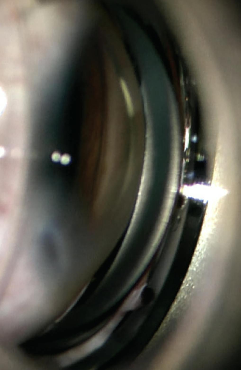
Figure 2. Postoperative gonioscopy reveals an open angle and well-pigmented trabecular meshwork from prior apposition to the iris.
One month after surgery, the patient reported no recurrence of his initial symptoms and a gradual improvement in his visual acuity. His BCVA was 20/80 OD and 20/25 OS. His unmedicated IOP was 16 mm Hg OD and 17 mm Hg OS.
The patient is scheduled to return yearly for a complete eye exam.
1. Chang TC, Tran KD, Cernichiaro-Espinosa LA, et al. Microcornea and thickened lens in angle closure following nonsurgical treatment of retinopathy of prematurity. J Ophthalmol. 2020;2020:7510903.
2. Michael AJ, Pesin SR, Katz LJ, Tasman WS. Management of late-onset angle-closure glaucoma associated with retinopathy of prematurity. Ophthalmology. 1991;98(7):1093-1098.
3. Fossum K, Knight TJ, Morrison DG, Joos KM. Lensectomy as treatment for refractory or progressive retinopathy of prematurity narrow-angle glaucoma. J AAPOS. 2024;28(2):103854.
4. Pollard ZF. Secondary angle-closure glaucoma in cicatricial retrolental fibroplasia. Am J Ophthalmol. 1980;89(5):651-653.
5. Mensher JH. Anterior chamber depth alteration after retinal photocoagulation. Arch Ophthalmol. 1977;95(1):113-116.
6. Ueda N, Ogino N. Angle-closure glaucoma with pupillary block mechanism in cicatricial retinopathy of prematurity. Ophthalmologica. 1988;196(1):15-18.
7. Trigler L, Weaver RG Jr, O’Neil JW, Barondes MJ, Freedman SF. Case series of angle-closure glaucoma after laser treatment for retinopathy of prematurity. J AAPOS. 2005;9(1):17-21.
8. Uehara A, Kurokawa T, Gotoh N, Yoshimura N, Tokushima T. Angle closure glaucoma after laser photocoagulation for retinopathy of prematurity. Br J Ophthalmol. 2004;88(8):1099-1100.
9. Lavanya R, Wong TY, Friedman DS, et al. Determinants of angle closure in older Singaporeans. Arch Ophthalmol. 2008;126(5):686-691.
10. Shah P, Bhakta A, Vanner EA, Kishor KS, Greenfield DS, Maharaj ASR. Safety and efficacy of diode laser transscleral cyclophotocoagulation in eyes with good visual acuity. J Glaucoma. 2018;27(10):874-879.


