Key Takeaways
- Myopia and unreliable visual field testing can make diagnosing glaucoma challenging.
- When standard perimetry is consistently unreliable, alternative strategies may be warranted.
- Genetic testing can provide insight into patients’ glaucoma risk.
CASE PRESENTATION
A 68-year-old woman presents for routine follow-up glaucoma care. She was initially referred by her optometrist 5 years ago because the appearance of the patient’s optic nerves raised suspicion of glaucoma, and she has been monitored as a glaucoma suspect ever since. The patient has no family history of the disease and no ocular complaints. She wears soft contact lenses for myopia and has been happy with her vision. She does not use eye drops.
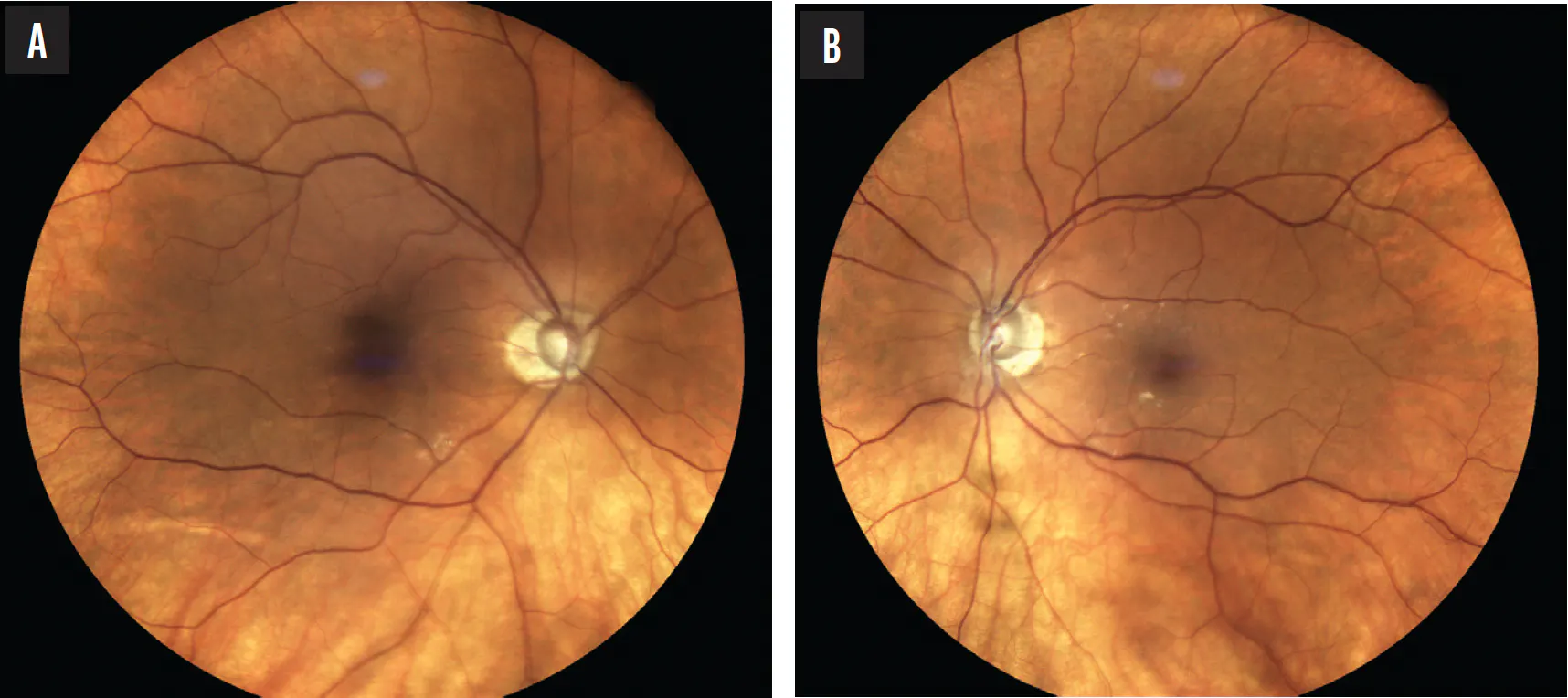
On examination, the patient’s BCVA is 20/20 OU, and a manifest refraction reveals -4.50 D of myopia in each eye. At her first visit, her IOP measured 16 mm Hg OU, but it has slowly risen since then. The current IOP reading is 19 mm Hg OU. Pachymetry shows a central corneal thickness of 492 µm OD and 490 µm OS. Gonioscopy demonstrates open angles to grade 4 in both eyes. A slit-lamp examination finds a 1+ nuclear sclerotic cataract in each eye. A fundus examination reveals a tilted nerve with peripapillary atrophy in each eye and cupping that is greater in the right eye (Figure 1).
Visual field testing (Humphrey Field Analyzer, Carl Zeiss Meditec) is unreliable with high false-positive errors in both eyes (Figure 2). This reflects a long-standing problem. Despite visual field coaching, the patient typically has very high false-positive errors. OCT reveals optic nerve cupping and thinning of the retinal nerve fiber layer (RNFL) that are worse in the right eye (Figure 3).
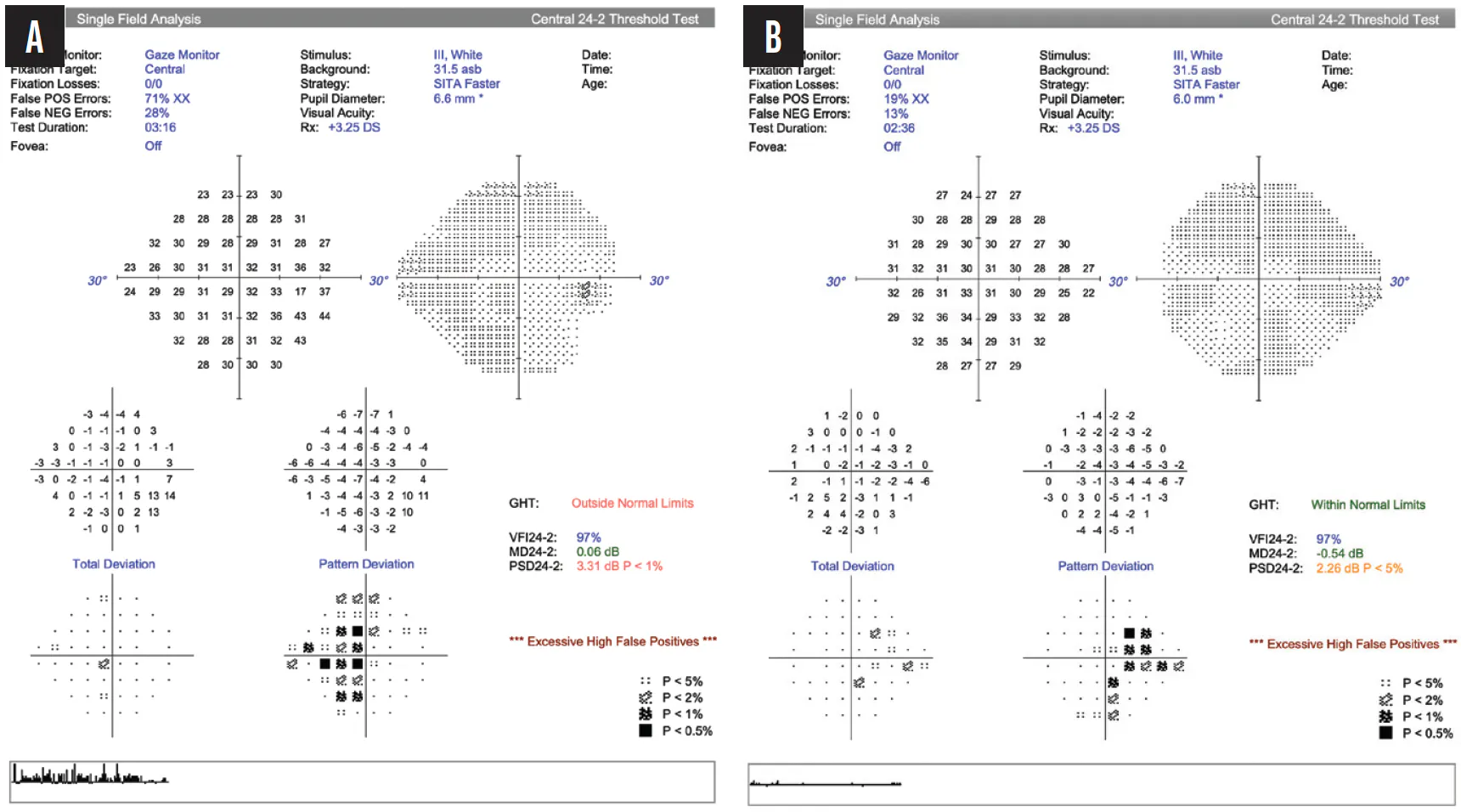
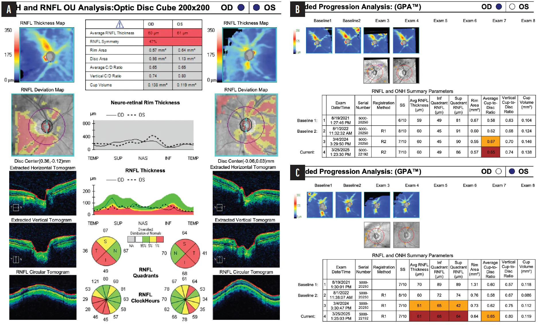
Figure 3. Most recent OCT scans of the optic nerve of each eye (A) and Guided Progression Analysis for the right (B) and left (C) eyes.
How would you address the patient’s difficulty with visual field testing? What is your interpretation of her OCT scans? Would you order other tests to assist with the risk stratification of this patient? How would you proceed with her glaucoma care?
—Case presentation by Jacob Brubaker, MD
JASON BACHARACH, MD
It is incumbent on glaucoma specialists to rule out masquerade syndrome before subjecting patients to a lifetime of treatment. Although pallor and swelling of the optic nerve do not appear to be evident, these masqueraders should be considered before suitable tools to monitor patients for disease progression are selected.
In this case, structural comparisons to the OCT machine’s normative database would be moot because of significant peripapillary atrophy. High false-positive rates would make the interpretation of functional testing with static automated perimetry using the Humphrey Field Analyzer challenging. The combination of rising IOP and thin pachymetry measurements is another concern.
The patient’s difficulty with visual field testing was not mitigated with coaching, and there was no improvement with repeat testing. Doing more of the same would therefore be unhelpful. Alternative visual field test-taking strategies such as the Goldmann kinetic test could be considered. Kinetic testing software, moreover, is embedded in some of the latest visual field technologies, and these systems do not require a highly skilled perimetrist to achieve accurate results.
OCT could help solidify the diagnosis, and Figure 3 suggests disease progression. The RNFL is thin overall, and it appears on assessment using the Guided Progression Analysis software (Carl Zeiss Meditec) to be thinning over time. An analysis of the ganglion cell complex could provide another structural marker less affected by the anomalous nature of the optic nerve head itself.
JEFFREY L. GOLDBERG, MD, PHD
In California, where I practice, I see a lot of glaucoma suspects with concomitant myopia. This patient’s open angles and 1+ nuclear sclerosis without other anterior segment anomalies reassure me that I am not missing another secondary glaucoma. I would not ascribe much significance to the increase in IOP from 16 to 19 mm Hg, even though corneal pachymetry suggests a slightly higher corrected IOP, because I try to avoid letting IOP influence diagnosis unless it is considerably higher (> mid-20s mm Hg).
The anatomic findings (eg, peripapillary atrophy), optic nerve cupping, and RNFL thinning together reflect how confounding it can be to differentiate myopia from early glaucoma. The RNFL thinning both superiorly and inferiorly and the increase in vertical cup-to-disc ratio observed over 3.5 years of OCT scans in both eyes may indicate glaucoma.
The inability to gather reliable visual fields is the biggest challenge in this situation, although reliable fields showing scotomas in either eye would not definitively differentiate glaucomatous from myopic degeneration. Documenting visual field progression in an older myopic adult would incline me toward a diagnosis of glaucoma. I would try Goldmann visual field testing because a useful result would help cement how highly to prioritize treatment.
In aggregate, the evidence points toward a presumptive diagnosis of glaucoma in addition to the patient’s mild myopia. Most important would be an honest discussion with her to clarify her priorities. Some patients’ greatest fear is starting treatment—even something as benign as selective laser trabeculoplasty (SLT) or monotherapy with a prostaglandin analogue—in which case offering observation with ongoing testing is a reasonable solution. Other patients’ greatest fear is losing vision from glaucoma, in which case offering initial therapy is very low risk. I would express to the patient that my preference would be to err on the side of treatment with SLT as a starting point and then offer regular follow-up and ongoing testing with OCT, including macular scans for ganglion cell complex tracking, which might not be subject to the same floor effects as the RNFL, and, ideally, Goldmann visual field testing.
CHRISTINE LARSEN, MD
Unfortunately, this situation is not atypical at a busy glaucoma clinic. High false-positive errors often make visual fields appear better than they really are and can distort global indices. The false-positive rate is one of the reliability parameters most associated with misleading visual field results. Although the reliability metrics for this patient limit the utility of the fields for progression analysis, the pattern deviation changes are not entirely random and may correlate loosely with the structural findings.
A trial of a size V stimulus might be helpful. Although not a validated default in individuals with 20/20 vision, this target can improve reproducibility and patients’ confidence during testing. Additionally, virtual reality perimetry is an emerging adjunctive modality that may improve the patient experience. I have had some success with this strategy in patients who have high false-positive rates; they often describe it as more of a game and less of a test.
It would be important not to overread so-called red disease on the RNFL printout, especially because the patient is myopic. Segmentation, scan centration, peripapillary atrophy, disc size, and longitudinal change would also be evaluated. High myopia increases the risk of an OCT artifact. Macular ganglion cell analysis and serial disc photography could therefore be particularly helpful. Although myopic tilt and peripapillary atrophy can confound RNFL interpretation, the longitudinal structural change and intereye asymmetry evident here could indicate true glaucomatous damage.
Given the patient’s thin pachymetry readings, increasing IOP, suspicious optic nerves, myopia, and OCT changes, intervention—likely primary SLT—would be reasonable. I would recommend treatment because her risk profile and structural findings are sufficient to justify therapy, even if functional confirmation is limited.
WHAT I DID: JACOB BRUBAKER, MD
Myopia, unreliable visual field tests—this case illustrates how difficult diagnosing glaucoma can be. The patient and I discussed how to improve test reliability several times to no avail. We also thoroughly discussed when and whether to consider therapy. I grew increasingly concerned about the patient’s care and proposed genetic testing in hopes it would provide insight into her situation.
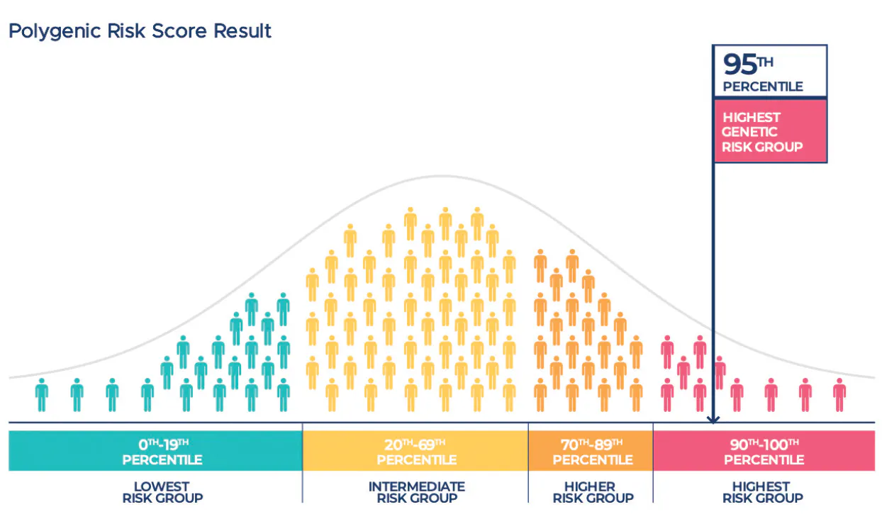
SightScore (Seonix Bio) uses a polygenic risk score to help determine an individual’s risk of developing glaucoma. Their genetic test results are compared to thousands of genetic variants known to be associated with glaucoma. Risk stratification is then provided based on these results.
A cheek swab was performed in the clinic. Four to 6 weeks after the sample was submitted, I received the patient’s results, which showed that she was in the highest-risk group (Figure 4). Given this information, she and I decided to initiate treatment.
After a discussion of the options, the patient elected to proceed with bilateral 360º manual SLT. Three weeks postoperatively, her IOP was 15 mm Hg OU.
The patient and I both felt that genetic testing was invaluable in guiding her care. She will continue to be closely monitored.