Key Takeaways
- Minimally invasive bleb surgery, or MIBS, has emerged as a distinct category within the MIGS spectrum to address a treatment gap for patients with glaucoma who have visual field loss and need enhanced IOP control but do not necessarily require extremely low IOPs.
- Two new technologies offer safer alternatives to traditional filtering procedures.
An Implant-Free Path to Minimally Invasive Subconjunctival Filtration
By Shan C. Lin, MD
MIGS has allowed glaucoma surgeons to treat early glaucoma, including ocular hypertension and mild to moderate disease, often in conjunction with cataract surgery. For more advanced disease, such as moderate to severe glaucoma, or in eyes with very high IOP, filtration surgeries are the most effective procedures to lower pressure and prevent progressive vision loss. However, trabeculectomy and tube shunt surgery have relatively high rates of complications, including hypotony, bleb leak, infection, and tube erosion. Minimally invasive bleb surgery (MIBS) has emerged as a distinct category within the MIGS spectrum to address a treatment gap for patients with glaucoma who have visual field loss and need enhanced IOP control but do not necessarily require extremely low IOPs.
A SAFER WAY TO ACHIEVE FILTRATION
MIBS encompasses procedures that achieve substantial IOP lowering through the creation of a subconjunctival filtering bleb via a microinvasive ab interno or ab externo approach. These procedures result in limited tissue disruption and demonstrate a safety profile that is comparable to that of angle-based MIGS.
The Aqualumen (PLU Ophthalmic) is a novel, implant-free surgical device that creates a controlled transscleral outflow pathway to reduce IOP in patients with glaucoma. The device features a modified 22-gauge needle with a back-cutting portion embedded within the shaft (Figure 1). The Aqualumen removes a core of scleral tissue as it is withdrawn from the eye to create filtration.
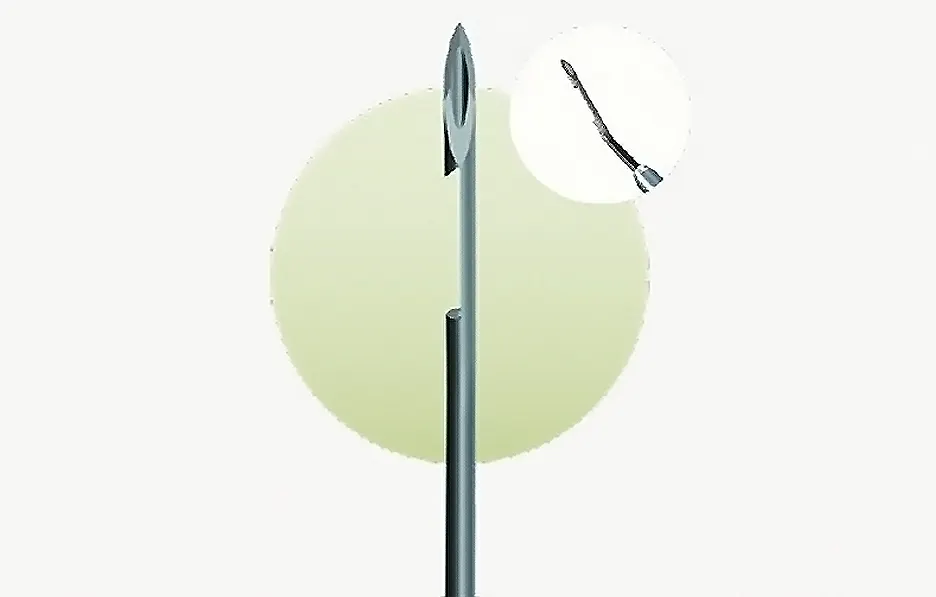
Figure 1. The Aqualumen device features a 22-gauge needle with a back-cutting blade embedded within the shaft to facilitate removal of a core of tissue during surgery.
By enabling diffuse, posterior bleb formation through a standardized, conjunctiva-sparing ab externo approach without scleral flap construction or a permanent implant, Aqualumen employs the principles of MIBS—combining the efficacy of traditional filtration surgery with a safety and recovery profile more typical of MIGS. This includes tissue preservation, a shorter operative time, and simplified postoperative management. By replicating the essential mechanism of trabeculectomy—direct aqueous filtration to the subconjunctival space—without a conjunctival opening or flap construction, Aqualumen is not limited by episcleral venous pressure.
PATIENT SELECTION
There is a wide spectrum of cases in which the Aqualumen procedure may be a useful approach to lower IOP and/or reduce the medication burden. Patients with existing visual field loss, those with uncontrolled IOP on multiple antiglaucoma drops, and those who experience significant side effects from medication may be candidates for MIBS procedures, including Aqualumen. Additionally, patients who wish to reduce their medication burden may benefit from the treatment because the IOP and number of medications are typically more significantly reduced after MIBS compared to MIGS.1
If a very low target IOP is necessary, such as in eyes with normal-tension glaucoma and a low baseline IOP or eyes with advanced glaucoma and disease progression despite a low IOP, trabeculectomy may be necessary to achieve an IOP of 10 mm Hg or lower. The Aqualumen procedure may be appropriate in some cases of secondary glaucoma, with the same caveats as in filtration surgery. Treatment success may be limited in eyes with angle-closure glaucoma owing to the higher risk for iris blockage of the inner opening compared with eyes with open-angle glaucoma. Similarly, the treatment may have a higher likelihood of failure in eyes with uveitic and neovascular glaucomas.
The conjunctiva-sparing nature of the Aqualumen procedure also preserves options for future surgery, which is especially valuable for patients who are younger or have advanced disease. Cases should be assessed individually, with careful consideration given to the patient’s clinical status, life expectancy, systemic conditions, and preferences.
SURGICAL TECHNIQUE AND PEARLS
A temporal paracentesis is created, and the anterior chamber is filled with an OVD. The Aqualumen device is then introduced into the subconjunctival space approximately 8 to 10 mm posterior to the limbus and advanced toward the limbal region. With firm globe fixation, the needle is directed through the sclera about 2 mm posterior to the limbus into the anterior chamber, entering at or just above the trabecular meshwork (Figure 2A).

Figure 2. A bird’s-eye view shows the orientation of the Aqualumen penetrating the anterior chamber (A). The device’s back-cutting blade engages the scleral tissue (B). The Aqualumen is removed with the excision of a scleral core, creating a tunnel for aqueous flow (C).
A rapid withdrawal of the device, while biasing the cutting edge against the scleral tissue, cores out a small tunnel and establishes the outflow pathway (Figure 2B and C). Additional passes can be made through the same entry if needed to optimize flow. The OVD is removed, and mitomycin C is injected subconjunctivally to modulate wound healing and support a posterior bleb.
Practical pearls based on early experience include the following:
- A generous amount of pre- and postoperative topical steroids should be administered to minimize conjunctival and Tenon fibrosis;
- Aqueous suppressants should be avoided at the conclusion of surgery; and
- A low threshold for postoperative needling should be maintained when early encapsulation is suspected.
Needling (passing the needle through the tunnel) is straightforward, helps ensure patency, and can clear any ostial obstruction.
EARLY STUDY RESULTS
A recent retrospective study from the Medical University of South Carolina reported early outcomes with the Aqualumen procedure in patients with refractory glaucoma.2 In 12 eyes of 12 patients, the most common glaucoma diagnosis was primary open-angle glaucoma (67%), followed by angle-closure glaucoma (17%), normal-tension glaucoma (8%), and uveitic glaucoma (8%). Baseline IOP among the entire cohort was 30.1 mm Hg. Patients who completed 6 months of follow-up achieved a 48.2% reduction in mean IOP, and half of them were free of all glaucoma medications. At the 12-month follow-up visit, the average IOP reduction was -14.5 mm Hg, and the mean medication reduction was -0.8.
Postoperative interventions were necessary in some cases, including needling in five eyes (38%), repeat Aqualumen in one eye (8%), and Ahmed Glaucoma Valve (New World Medical) implantation in one eye (8%). The greatest risk factor for additional intervention was combination with cataract surgery. All five eyes that underwent the Aqualumen procedure combined with cataract surgery required postoperative intervention, compared with one eye in the standalone group that required repeat treatment with Aqualumen.
SAFETY PROFILE AND COMPLICATIONS
In the retrospective study, the most common adverse events were cystoid macular edema (33%), ostium obstruction by the iris (25%), bleb fibrosis (17%), and transient corneal edema (8%). There were no cases of hypotony or vision-threatening side effects, and all cases of cystoid macular edema resolved with treatment. Serious complications typically associated with trabeculectomy, such as chronic hypotony, endophthalmitis, and late bleb leak, were not observed in this data set. Nevertheless, the small sample size and relatively short follow-up period warrant cautious interpretation. Larger, long-term studies are required to define the Aqualumen’s extended safety profile.
CONCLUSION
New and effective alternatives to traditional filtration surgeries are welcome developments given the high risks associated with trabeculectomy and tube shunt surgery. MIGS procedures have become more common in recent years, but they are indicated for ocular hypertension and milder stages of glaucoma.3 The emergence of MIBS, including the Aqualumen procedure, addresses this gap in glaucoma care. Early evidence for the Aqualumen shows effective IOP and medication reductions without a significant risk of serious, vision-threatening adverse events. For eyes with moderate to severe glaucoma, the Aqualumen offers an implant-free surgical option that has the potential to achieve safe and effective aqueous filtration.
1. Konopińska J, Gołaszewska K, Saeed E. Minimally invasive bleb surgery versus minimally invasive glaucoma surgery: a 12-month retrospective study. Sci Rep. 2024;14(1):12850. doi:10.1038/s41598-024-61811-y
2. Irfan MA, Hindi A, An J. Early outcomes of a novel minimally invasive filtering surgery in refractory glaucoma: a pilot study. Poster presented at: American Glaucoma Society Annual Meeting; February 19-22, 2026; Rancho Mirage, CA.
3. Lin SC, Lum F, Liu L, et al. Outcomes of minimally invasive and traditional incisional glaucoma surgery: an IRIS (Intelligent Research In Sight) Registry study. AJO International. 2026;3:100246. doi.org/10.1016/j.ajoint.2026.100246
office-based intervention: glaucoma surgery at the Slit Lamp
By Nir Shoham-Hazon, MD
The glaucoma armamentarium features a variety of MIGS procedures that help bridge the gap between noninvasive first-line therapies such as medications and laser ablation and invasive filtration surgery for refractory or advanced disease. This paradigm change has been driven by an increasingly widespread understanding of the benefits of interventional glaucoma care, such as avoiding the challenges of patient adherence to medical therapy, ocular surface disease, and the costs of long-term care.1
Most MIGS procedures are reserved for patients with mild to moderate glaucoma because the IOP reduction from trabecular outflow–based procedures is modest due to a floor effect caused by episcleral venous pressure.2 This issue is bypassed by a specific type of MIGS that shunts aqueous humor into the subconjunctival space, similar to traditional filtration surgeries such as trabeculectomy. These MIBS procedures can achieve a greater IOP reduction than other MIGS procedures2 and, in some studies, have demonstrated efficacy similar to that of traditional filtration surgery.3
OPTIMIZING MIBS
Despite their advantages, current MIBS and MIGS procedures are performed exclusively in the OR, resulting in two significant limitations. The first is increased costs associated with the infrastructure and staff requirements, which reduces patient access to these procedures in resource-limited settings. The second is patient distress caused by the wait times, hospital admission, and fear of surgery.4
The office-based intervention (OBI) glaucoma device, OBI Core (Hexiris Ophthalmics), is an intuitive scleral punch system with a 27-gauge needle that is 6 mm long. The needle is loaded on a handpiece with an actuator for advancement into the anterior chamber (Figure 3). An oblique, posteriorly directed scleral microtunnel (diameter, 150 ±50 µm) is created that shunts aqueous humor from the anterior chamber to a posterior sub-Tenon bleb (Figure 4). The length of the microtunnel provides intrinsic hydraulic resistance for the controlled microflow of aqueous humor, reducing the risk of early hypotony until bleb healing is established. Unlike other MIBS procedures, an OBI Core sclerostomy trephination can be performed either as a rapid standalone office-based procedure at the slit lamp or in combination with phacoemulsification in the OR. This versatility has the potential to enhance the accessibility and convenience of interventional MIBS in diverse practice settings, particularly in regions where patient access to glaucoma procedures is limited.4,5
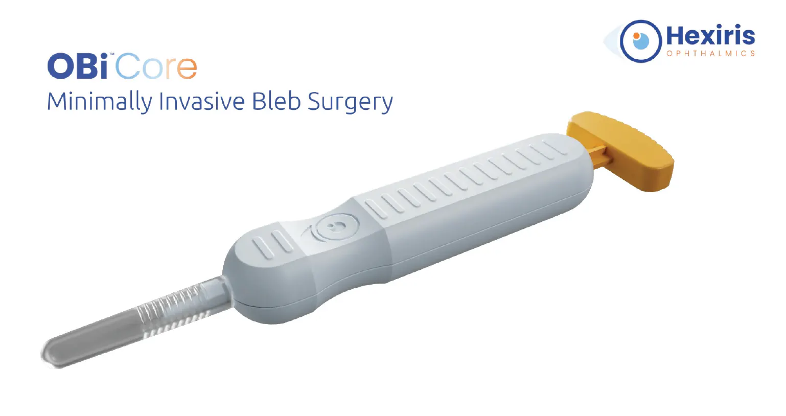
Figure 3. The 27-gauge needle of the OBI Core device is 6 mm long and loaded on a handpiece with an actuator for needle advancement into the anterior chamber.
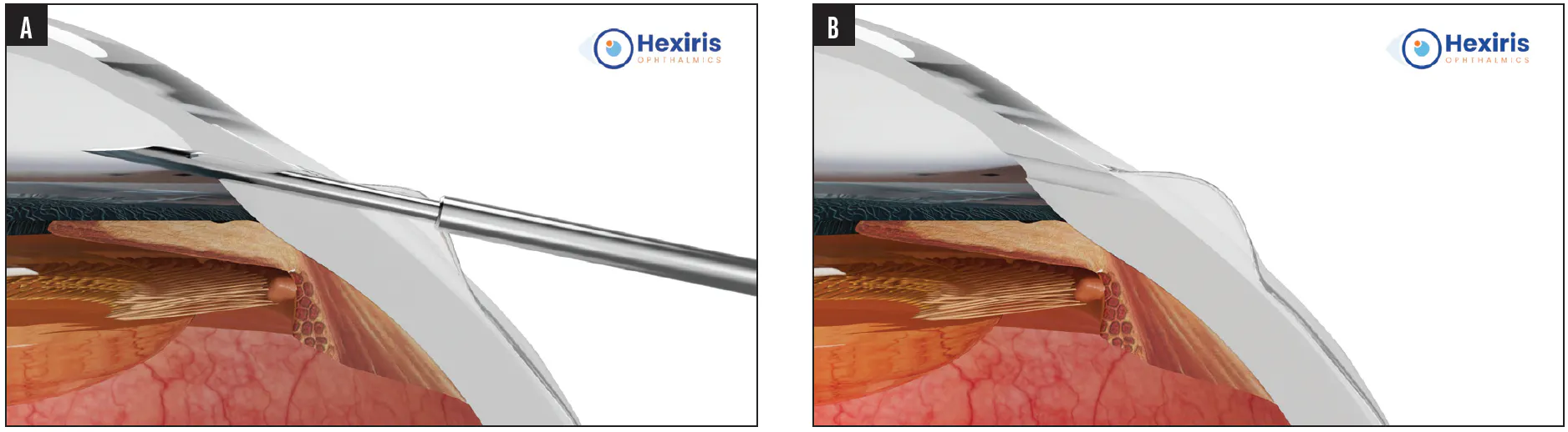
Figure 4. Punch excision of the sclera is performed with the needle of the OBI Core device directed into the anterior chamber using an ab externo approach (A). An oblique scleral microtunnel connecting the anterior chamber to a posterior sub-Tenon bleb is created (B).
PATIENT SELECTION
The OBI Core is recommended both as a primary procedure and as a reintervention in patients with moderate to advanced open-angle glaucoma whose IOP is uncontrolled on maximal tolerated medical therapy and in those who are intolerant of topical drop therapy or struggle with adherence. The procedure can also be appropriate when surgical intervention is not feasible because of prior failed filtration surgery or the patient’s preference.
SURGICAL TECHNIQUE
Controlled subconjunctival hydrodissection is performed in the superotemporal quadrant using a mixture of lidocaine and epinephrine (0.1 mL) and balanced salt solution (0.1–0.2 mL) injected through a 27-gauge needle (Figure 5). Some of the injected volume is directed toward the planned microtunnel ostium to prevent postoperative tissue apposition and obstruction, but the majority is aimed posteriorly to create a broad, smooth pocket 6 to 8 mm posterior to the limbus that promotes diffuse posterior filtration.
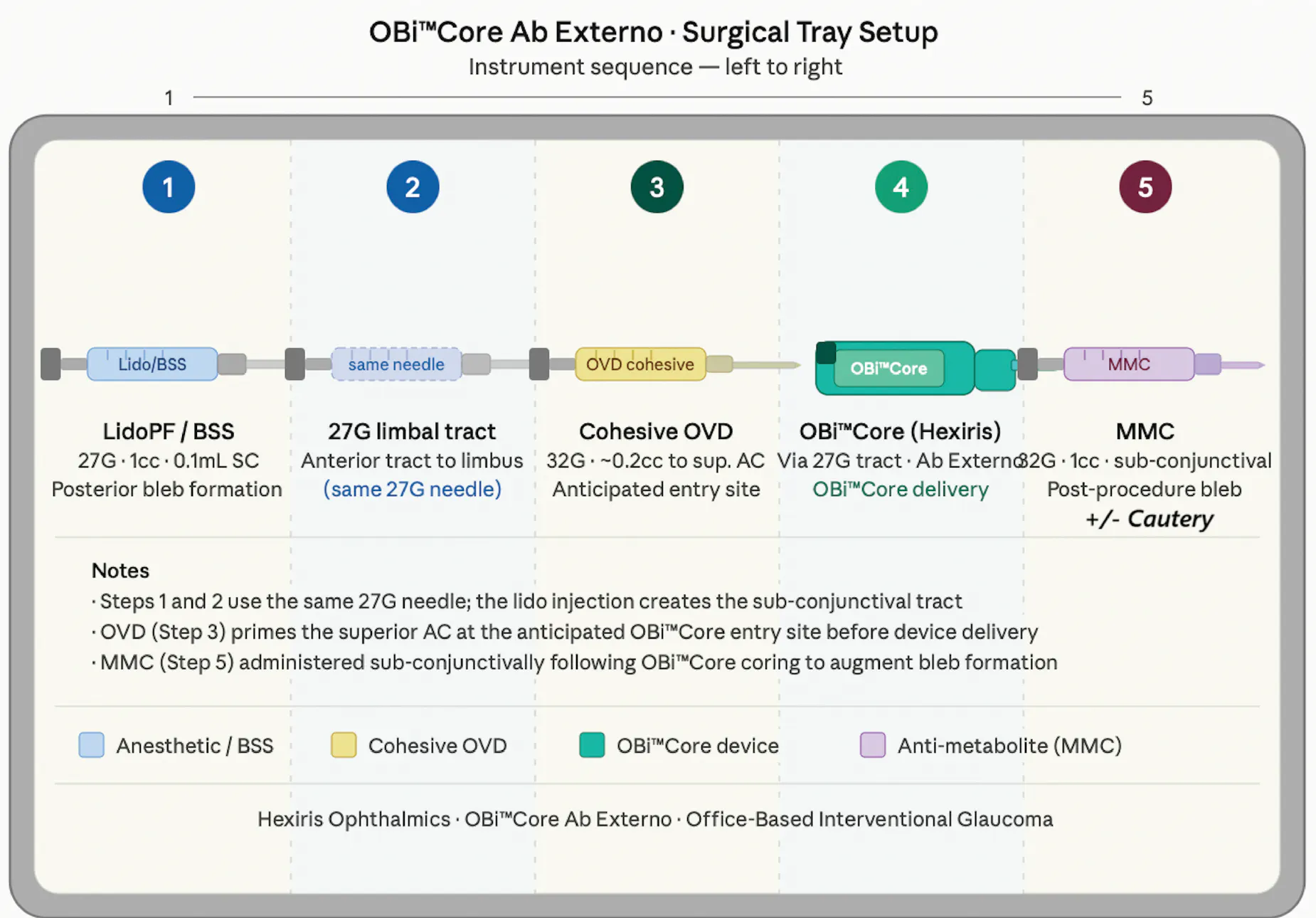
Figure 5. Schematic surgical tray setup for the OBI Core procedure. Instruments are arranged from left to right in order of use.
Next, a cohesive OVD bolus is placed in the anterior chamber to limit early postoperative microflow and reduce the risk of hypotony. The OBI Core is then placed against the superotemporal sclera, 1.0 to 1.5 mm posterior to the limbus. The actuator is depressed with the device positioned so that the punch excision is oriented anteriorly or slightly laterally to avoid iris incarceration and encourage diffuse bleb formation. Mitomycin C (0.4 mg/mL) is injected into the previously created subconjunctival pocket to augment bleb formation. At the end of the case, pilocarpine 1% is administered to pull the iris away from the ostium. A Seidel test is performed; if it is positive, gentle conjunctival cautery is typically sufficient.
Postoperative care includes regular monitoring of the bleb’s appearance and IOP. If the IOP is elevated (> 21 mm Hg) with minimal bleb formation, gentle digital massage can help encourage filtration through the ostium. Needling can be considered after the first postoperative week if filtration is reduced by early subconjunctival fibrosis. Hypotony (IOP < 5 mm Hg) with a shallow anterior chamber can be managed with an injection of a cohesive OVD at the slit lamp. In the rare event of iris incarceration, miotic therapy should be initiated. Laser iridoplasty/iridotomy may be considered if necessary.
CLINICAL OUTCOMES
The OBI Core’s first-in-human outcomes were presented at the 2026 ASCRS Annual Meeting.6 In a preliminary cohort of 10 eyes (10 patients) monitored for up to 6 weeks after standalone office-based OBI Core surgery, the IOP was reduced from 30.1 ±9.0 to 10.2 ±7.2 mm Hg, and the number of glaucoma medications decreased from 2.2 ±1.7 to 0.8 ±1.6, with mean reductions of 19.9 mm Hg and 1.4 medications. The results were consistent across the cohort, with 70% of eyes achieving complete success (defined as an unmedicated IOP < 21 mm Hg) and 78% of eyes becoming medication free.
Patients experienced no significant change in their visual acuity from baseline to the last follow-up visit (0.59 ±0.66 to 1.08 ±0.70 logMAR), and none experienced vision loss. Although no intra- or postoperative complications occurred, 30% of eyes required reintervention, which included either repeat OBI Core procedures or other filtration procedures.
CONCLUSION
The Hexiris OBI Core is designed to optimize the accessibility and convenience of MIBS while retaining its IOP-lowering benefits. The procedure allows a new class of interventions that can be performed either at the slit lamp or in the OR. First-in-human results with standalone office-based OBI Core safely achieved postoperative IOPs similar to those obtained with trabeculectomy. The aim of an ongoing multicenter case series with a longer follow-up period is to validate the initial findings, and associated cost-effectiveness analyses will quantify the potential cost savings of one-time OBI Core intervention. Growing clinical experience is expected to define the role of the device in the interventional glaucoma care continuum.
1. Funke CM, Ristvedt D, Yadgarov A, Micheletti JM. Interventional glaucoma consensus treatment protocol. Expert Rev Ophthalmol. 2025;20(2):79-87. doi:10.1080/17469899.2025.2465330
2. Gillmann K, Mansouri K. Minimally invasive glaucoma surgery: Where is the evidence? Asia Pac J Ophthalmol (Phila). 2020;9(3):203-214. doi:10.1097/APO.0000000000000294
3. Traverso CE, Carassa RG, Fea AM, et al. Effectiveness and safety of Xen Gel Stent in glaucoma surgery: a systematic review of the literature. J Clin Med. 202312(16):5339. doi:10.3390/jcm12165339
4. Sarfo JO, Mordi P, Aggrey EK, Quaicoe ASP, Attafuah PYA. Glaucoma prevalence and treatment in Sub-Saharan Africa’s elderly population: a scoping review. BMC Geriatr. 202525(1):255. doi:10.1186/s12877-025-05901-0
5. Ramesh PV, Ray P, Senthil Kumar NK, Ramesh SV, Devadas AK. Commentary: minimally invasive glaucoma surgery for a surgical take diversion: an economic perspective. Indian J Ophthalmol. 2023;71(2):566-568. doi:10.4103/ijo.IJO_2264_22
6. Harasymowycz P, Ahmed I, Gagne S. Office-based slit-lamp interventional glaucoma surgery: early first-in-human clinical outcomes and preliminary economic rationale. Paper presented at: ASCRS Annual Meeting; April 10-13, 2026; Washington, DC.