


Key Takeaways
- Angle closure is best conceptualized not as a single diagnosis but as a spectrum.
- The staging of angle closure matters clinically because it reframes management away from laser peripheral iridotomy versus lensectomy as a binary decision and toward a more nuanced calculus
- Key considerations include the patient’s disease stage, age, and presbyopic status.
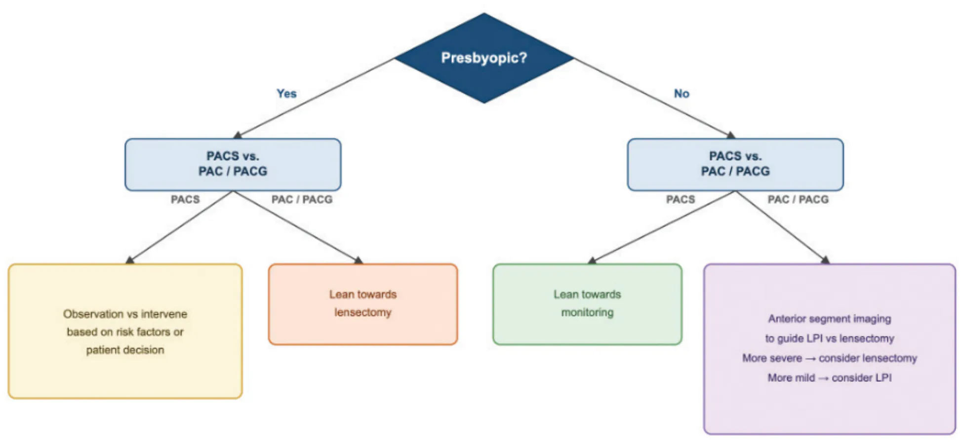
Angle closure is best conceptualized not as a single diagnosis but as a spectrum. The term primary angle-closure suspect (PACS) describes appositional anatomy alone. PAC presents with either elevated IOP or peripheral anterior synechiae (PAS) without evidence of optic nerve damage. The term primary angle-closure glaucoma (PACG) refers to the stage at which optic neuropathy has occurred. This staging matters clinically because it reframes management away from laser peripheral iridotomy (LPI) versus lensectomy as a binary decision and toward a more nuanced calculus (Figure). The questions become the following: Where does this eye sit on the spectrum, and what does the patient’s age, and more specifically, their presbyopic status, demand of the conversation?
THE PACS PATIENT
Regardless of their age, for patients with appositional angles alone and no PAS, IOP elevation, or evidence of nerve damage, management is a discussion rather than an algorithm. Risk factors, fellow-eye and family history, the logistic burden of frequent dilated exams, access to care, and symptoms suggestive of intermittent angle closure all factor into whether a patient would be better served by observation, LPI, or early lens extraction. Both the Zhongshan Angle-Closure Prevention (ZAP) trial and the Singapore Asymptomatic Narrow Angles Laser Iridotomy Study (ANA-LIS) demonstrated that LPI reduced the risk of progression to PAC or PACG, but the absolute event rate in untreated PACS eyes was quite low1,2; the number needed to treat is high enough that a prophylactic LPI cannot be justified for all PACS patients.1,2
THE PRESBYOPIC PATIENT
The calculus changes considerably for a patient with presbyopia. The conversation becomes simpler, because the principal cost of lensectomy—the loss of accommodation—is no longer a major factor. The Effectiveness of Early Lens Extraction for the Treatment of Primary Angle-Closure Glaucoma (EAGLE) trial supported this approach. The study showed that clear lens extraction was often preferable to LPI in PAC eyes with an IOP of 30 mm Hg or higher or in PACG eyes.3 However, we do not recommend waiting for the IOP to reach 30 mm Hg or any other specific numeric target before acting. If the IOP is consistently above normal or if optic nerve damage has occurred, surgery is warranted on a presbyopic eye.
THE PREPRESBYOPIC PATIENT
The bar for lensectomy rises meaningfully in eyes with PAC or PACG that retain accommodative ability. Anterior segment imaging, whether ultrasound biomicroscopy or anterior segment OCT, is useful to characterize the mechanism of angle closure. If pupillary block is the predominant mechanism in an eye with PAC or PACG, LPI remains a rational first step. If the mechanism is not primarily related to pupillary block, such as plateau iris, other interventions must be considered. The angle findings themselves are informative: Eyes with more extensive PAS are more likely to require a lensectomy, whereas eyes with predominantly appositional closure and minimal synechial change may be more amenable to an LPI.
Nonetheless, when a younger patient elects to undergo a lensectomy, the conversation must explicitly address presbyopia and the loss of accommodation. Presbyopia-correcting IOLs are often an attractive option in this situation because angle closure differs mechanistically from progressive optic neuropathies such as primary open-angle glaucoma, pseudoexfoliative glaucoma, and pigmentary glaucoma. Once the anatomic driver of angle closure is removed via lensectomy, further disease progression is not expected, unlike with other glaucomatous etiologies. This is an important distinction when counseling a young patient about the long-term suitability of a presbyopia-correcting lens that can help them see at multiple distances.
“The task is to match the timing and extent of surgery to where the eye sits on the angle-closure continuum, to the patient’s accommodative status, and to their individual risk profile and preferences instead of applying a single protocol to a disease that is anything but uniform.”
BIOMETRIC CONSIDERATIONS
For most eyes with an axial length (AL) greater than 21 mm, the Barrett Universal II formula performs well. For an AL of less than 21 mm, triangulating across multiple formulas may be more successful than relying on a single one. An eye with relative anterior microphthalmos deserves particular caution: Modern formulas use effective lens position, an assumption that becomes unreliable when the anterior segment is short but the length of the posterior segment is normal. In these eyes, defaulting to an older-generation formula such as Holladay 1 may be helpful.4 In this setting, we recommend aiming for a slightly hyperopic outcome because the IOL may sit more anteriorly than modern effective lens position modeling predicts, which, if not accounted for, would induce a myopic refractive error.
ADJUNCTIVE PROCEDURES
Several concurrent interventions merit consideration alongside lensectomy for patients with angle closure. Goniosynechialysis is a low-risk, reasonable option when PAS are present. We believe that restoring normal anatomy is the best approach to ensure long-term outflow facility and thus clinical stability. Endocyclophotocoagulation can be useful specifically in plateau iris configuration, where shrinking the ciliary processes helps deepen the angle.
In eyes with an AL of less than 18 mm, prophylactic scleral windows deserve consideration. A randomized controlled trial demonstrated that prophylactic sclerostomy performed at the time of cataract surgery in nanophthalmic eyes reduced complication rates, particularly of uveal effusion, in a population otherwise prone to vision-threatening intra- and postoperative events.5 Concurrent iridozonulohyaloidovitrectomy can be reserved for eyes with an AL of less than 20 mm or a fellow-eye history of malignant glaucoma. Canal-based MIGS, goniotomy, off-label microstenting, and canaloplasty may be considered in eyes with more severe disease or a high medication burden, although the evidence supporting these approaches remains limited.
Lensectomy is the primary intervention across much of this spectrum, but that does not mean it must be performed immediately for all patients. The task is to match the timing and extent of surgery to where the eye sits on the angle-closure continuum, to the patient’s accommodative status, and to their individual risk profile and preferences instead of applying a single protocol to a disease that is anything but uniform.
Authors’ AI disclosure: An AI language model (Claude Sonnet 5, Anthropic) was used to assist in the creation of the figure. All content was reviewed, verified, and revised by the authors, who assume full responsibility for the accuracy and integrity of the manuscript. The AI tool was not used for data analysis, interpretation, or drawing scientific conclusions.
1. He M, Jiang Y, Huang S, et al. Laser peripheral iridotomy for the prevention of angle closure: a single-centre, randomised controlled trial. Lancet. 2019;393(10181):1609-1618. doi:10.1016/S0140-6736(18)32607-2
2. Baskaran M, Kumar RS, Friedman DS, et al. The Singapore Asymptomatic Narrow Angles Laser Iridotomy Study: five-year results of a randomized controlled trial. Ophthalmology. 2022;129(2):147-158. doi:10.1016/j.ophtha.2021.08.017
3. Azuara-Blanco A, Burr J, Ramsay C, et al. Effectiveness of Early Lens Extraction for the Treatment of Primary Angle-Closure Glaucoma (EAGLE): a randomised controlled trial. Lancet. 2016;388(10052):1389-1397. doi:10.1016/S0140-6736(16)30956-4
4. Jung KI, Yang JW, Lee YC, Kim SY. Cataract surgery in eyes with nanophthalmos and relative anterior microphthalmos. Am J Ophthalmol. 2012;153(6):1161-1168.e1. doi:10.1016/j.ajo.2011.12.006
5. Rajendrababu S, Babu N, Sinha S, et al. A randomized controlled trial comparing outcomes of cataract surgery in nanophthalmos with and without prophylactic sclerostomy. Am J Ophthalmol. 2017;183:125-133. doi:10.1016/j.ajo.2017.09.008


