


Key Takeaways
- Supraciliary glaucoma surgery augments uveoscleral outflow through an ab interno clear corneal microincision and is frequently combined with phacoemulsification.
- Potential refractive errors may be a transient change from early hypotony or a sustained shift mediated by axial length changes.
- Because supraciliary surgery uses no scleral flaps, sutures, or bleb, surgically induced astigmatism tracks the corneal incision rather than the implant.
Supraciliary glaucoma surgery augments uveoscleral outflow through an ab interno clear corneal microincision and is frequently combined with phacoemulsification. Potential refractive errors may be caused by two distinct processes: (1) a transient change from early hypotony and (2) a sustained shift mediated by axial length (AL) changes. Because supraciliary surgery uses no scleral flaps, sutures, or bleb, surgically induced astigmatism tracks the corneal incision rather than the implant. The effect is akin to that of microincisional cataract surgery alone if the wound is constructed in a similar fashion.1
CASE PRESENTATION
A patient with mild to moderate primary open-angle glaucoma on three topical medications presented with a visually significant cataract and an IOP of approximately 24 mm Hg.
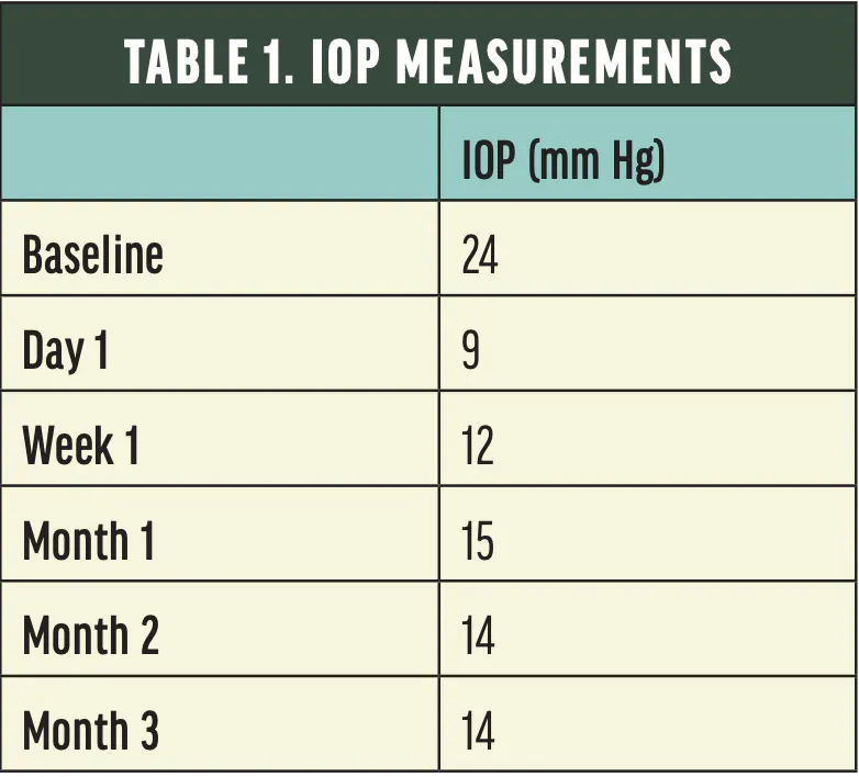
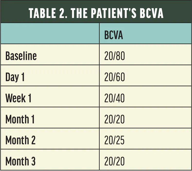
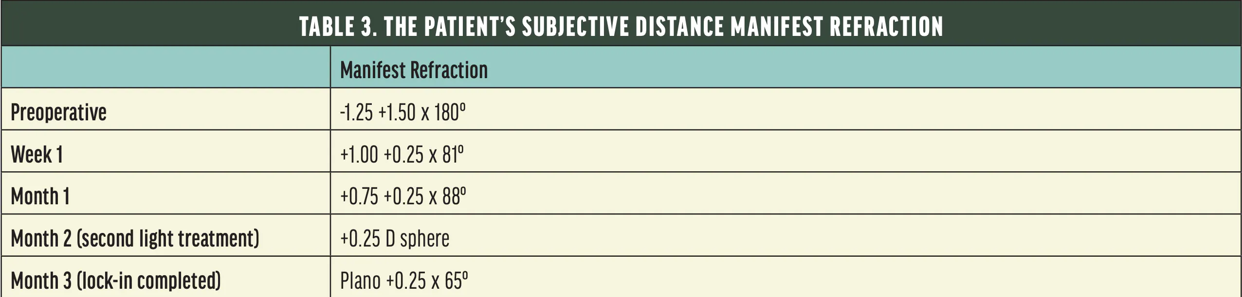
The patient underwent uncomplicated phacoemulsification through a temporal clear corneal microincision and placement of a Light Adjustable Lens (LAL; RxSight) in the bag, followed by ab interno supraciliary outflow enhancement with bio-reinforcement using the AlloFlo system (Iantrek). Their visual acuity, IOP, and refraction with spherical equivalent were monitored for 3 months (Tables 1–3).
Postoperatively, the IOP decreased to 15 mm Hg. As the IOP normalized, a modest hyperopic shift in spherical equivalent emerged that was consistent with AL shortening, but the amount of refractive cylinder was essentially unchanged. A slight, transient hyperopic shift occurred during the first week of low IOP and persisted through postoperative month 1. In response, light treatments were performed to fine-tune the LAL toward an emmetropic result.2 The patient’s target visual acuity was achieved 1 month after surgery. Their refraction was slightly more hyperopic than the target, a difference attributable to the postdecompression AL change, and this discrepancy was also addressed with a light treatment of the LAL.
DISCUSSION
With supraciliary glaucoma procedures, surgically induced astigmatism is driven by the incision, not the glaucoma device. A soft, porous supraciliary implant should not add a measurable amount of astigmatism.1,3,4
A common concern regarding the use of supraciliary devices is the potential myopic shift, as was observed with the CyPass MicroStent (Alcon; no longer available). An advantage of the LAL is the ability to make refractive adjustments postoperatively—in this case, to address an IOP-dependent hyperopic shift.
The timing of biometry affects its predictability. Capturing measurements at elevated IOPs increases the risk of a hyperopic surprise.5-9
Early hypotony temporarily degrades visual quality. The condition peaks within a week of surgery and typically resolves by approximately 2 months. The final refraction should be deferred until the IOP and corneal shape stabilize.2
The predicable but variable hyperopic drift that accompanies a sustained IOP reduction has long been the hidden cost of combined glaucoma and cataract surgery. With appropriate planning and counseling, patients can have highly successful outcomes.
1. Nakagawa S, Kato Y, Totsuka K, Kanda S, Okinaga K, Ishii K. Surgically induced astigmatism and refractive outcomes after minimally invasive glaucoma surgery combined with cataract surgery. Sci Rep. 2025;15(1):13966. doi:10.1038/s41598-025-96619-x
2. Baba T, Hirooka K, Okada N, et al. Changes in corneal higher-order aberrations following PreserFlo MicroShunt implantation. Sci Rep. 2025;15(1):16748. doi:10.1038/s41598-025-01550-w
3. Englisch CN, Wakili P, Langenbucher A, et al. Refractive and corneal astigmatism after implantation of a supraciliary drainage device in microinvasive glaucoma surgery. J Refract Surg. 2026;42(6):e565-e571. doi:10.3928/1081597X-20260413-01
4. Englisch CN, Boden KT, Messias A, et al. Short-term refractive and corneal astigmatism after canal-based microinvasive glaucoma surgery. PLoS One. 2026;21(1):e0340377. doi:10.1371/journal.pone.0340377
5. Kosaka Y, Kojima T, Tamaoki A, et al. Impact of axial length correction in high intraocular pressure eyes on intraocular lens power calculation. PLoS One. 2026;21(5):e0349117. doi:10.1371/journal.pone.0349117
6. Chirapapaisan C, Eiamsamarng A, Chirapapaisan N, et al. Effects of intraocular pressure change on intraocular lens power calculation in primary open-angle glaucoma and ocular hypertension. PLoS One. 2024;19(6):e0304169. doi:10.1371/journal.pone.0304169
7. de Sousa Franco CGV, Akio Pereira IE, Pereira ACA, et al. Refractive outcomes and predictability after cataract surgery combined with GATT or Kahook Dual Blade goniotomy. Sci Rep. 2026;16(1):7137. doi:10.1038/s41598-026-38240-0
8. Nakagawa S, Kaburaki T, Ishii K. Should Schlemm canal-based MIGS be combined with cataract surgery in patients receiving topical glaucoma therapy? A cataract surgeon-oriented review. J Clin Med. 2026;15(14):5503. doi:10.3390/jcm15145503
9. Saeedi O, Pillar A, Jefferys J, Arora K, Friedman D, Quigley H. Change in choroidal thickness and axial length with change in intraocular pressure after trabeculectomy. Br J Ophthalmol. 2014;98(7):976-979. doi:10.1136/bjophthalmol-2013-304433

