Key Takeaways
- Taking a patient to the OR for a controlled revision procedure demands a significant amount of time and resources.
- If a slit-lamp procedure might safely spare a patient a trip to the OR, it is generally worth attempting.
- Slit-lamp procedures glaucoma surgeons should know include anterior chamber tap, needling, tube sweep, and hyphema drainage.
Taking a patient to the OR for a controlled revision procedure holds undeniable appeal, but it also demands a significant amount of time and resources. Urgent additions to an already full surgical schedule often delay or displace other cases. Estimates of OR costs range from approximately $500 to $1,000 per hour in ambulatory surgery centers to $2,000 to $3,000 per hour in hospital-based ORs.1,2 If a slit-lamp procedure might safely spare a patient a trip to the OR, it is generally worth attempting.
This article discusses several slit-lamp procedures that every glaucoma surgeon should have in their armamentarium. For all the techniques, the ocular surface may be sterilized with topical antibiotic drops before and after the procedure or with topical povidone-iodine. Re-formation of the anterior chamber (AC) with an OVD is not discussed, as most glaucoma surgeons are familiar with this technique and its utility.
AC TAP
Practice patterns vary greatly among institutions, but during my (I.O.) training, it was uncommon to go more than a week without performing at least one AC tap. Even in glaucoma practice, where I rarely send aqueous samples for viral polymerase chain reaction testing, AC taps remain a valuable therapeutic and diagnostic procedure.
My (I.O.) technique is straightforward and uses a 30-gauge needle attached to a 1-mL syringe with the plunger completely withdrawn. After the eye has been anesthetized with topical proparacaine, a cotton-tipped applicator soaked in anesthetic is applied to the inferotemporal cornea for approximately 15 seconds. The needle’s passage into the AC has a slightly downward trajectory to avoid creating an overly long corneal tunnel. Typically, no countertraction is necessary. Care is taken to avoid the iris and crystalline lens. Once the needle is inside the AC, aqueous is allowed to egress slowly for approximately 10 seconds, and then, the AC is gently decompressed while the needle is withdrawn. In my experience, even a postprocedural IOP of 3 mm Hg is generally well tolerated provided the AC does not flatten.
From a therapeutic standpoint, AC taps can provide dramatic relief to patients experiencing headache, pain, and corneal edema from acutely elevated IOP. Topical medications often penetrate an edematous cornea poorly, and reducing the IOP can restore the eye’s ability to respond to medical therapy. Much like blowing into an old Nintendo cartridge, occasionally an AC tap is all that is needed to lower a patient’s IOP to within a manageable range.
The diagnostic utility of AC taps is perhaps underappreciated. After a goniotomy, it may be unclear how well the conventional outflow pathway is functioning or whether the procedure adequately unroofed the trabecular meshwork. If an AC tap lowers the IOP below episcleral venous pressure (approximately 8–10 mm Hg),3 blood reflux from Schlemm canal should occur in an eye with a functioning outflow system. In patients with a suboptimal response to angle surgery, the absence of heme reflux may reveal scarring or incomplete treatment. Repeating a goniotomy may allow a patient to avoid filtering surgery.
NEEDLING PROCEDURES
Needling encompasses a variety of techniques involving the introduction of a needle into the subconjunctival space following glaucoma surgery. Effective needling can resurrect a seemingly nonfunctional bleb after a trabeculectomy or the placement of a Xen Gel Stent (AbbVie) and spare the patient a return trip to the OR. Similarly, the decompression of an encapsulated bleb around the plate of a glaucoma drainage device may improve the patient’s level of comfort and long-term bleb function.
Lidocaine jelly is applied to the surface and allowed to remain in place for a few minutes. A 30-gauge needle is passed into the subconjunctival space, well away from the bleb, and a superficial plane is maintained. Careful dissection of the scarred Tenon tissue is performed. Depending on the degree of IOP elevation, either the scleral flap is gently elevated, or entrance into the anterior chamber is completed through the flap if necessary. Mitomycin C (MMC) or 5-fluorouracil is injected posterior to the bleb as needed. These procedures can be uncomfortable for patients when performed at the slit lamp, and some of them ultimately require treatment in the OR.
A similar approach may be used for Xen needling. Scar tissue above and below the implant is released to ensure it is mobile (Figure 1). The bleb is then entered, and further adhesions are broken. This tactile procedure allows the surgeon to feel any fibrotic bands in the bleb. An OVD may be injected into the bleb to expand it further. If a sufficient amount of time has elapsed since surgery and the bleb does not exhibit significant avascularity, I (I.O.) often inject 0.05 to 0.1 mL of 40 mg/mL MMC into the superior subconjunctival space and allow it to diffuse for approximately 10 minutes before proceeding.4
Overencapsulation occurs most commonly with valved drainage devices, young patients, and eyes with neovascular glaucoma.5 For encapsulated tube blebs, I (I.O.) prefer a large needle, typically 27- or 25-gauge, attached to a 3-mL syringe (Figure 2). Entering the bleb can be more difficult than anticipated because of dense fibrosis. I gently sweep the needle to break adhesions over the plate and then aspirate fluid from the bleb cavity. It is not uncommon for more than 1 mL of aqueous to be removed from a large, encapsulated bleb.
Physiologically, blebs tend to become encapsulated in response to increased wall tension.6 After the bleb has been decompressed and aqueous suppression has been initiated, patients often report that the bleb feels smaller and more comfortable at follow-up. Decompression may be repeated every 1 to 2 weeks if a bleb remains significantly enlarged. Some physicians further enhance bleb decompression with the injection of an antifibrotic agent such as 5-fluorouracil or MMC. If the capsule feels very thick, it is likely that a capsulectomy in the OR would be beneficial.
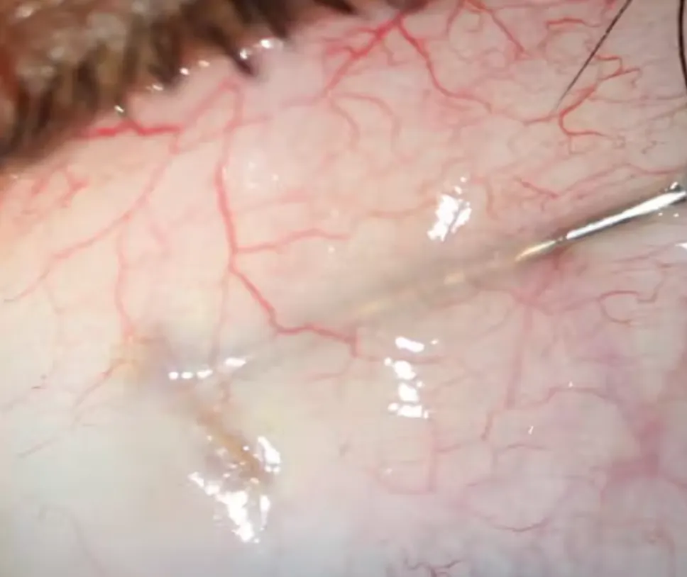
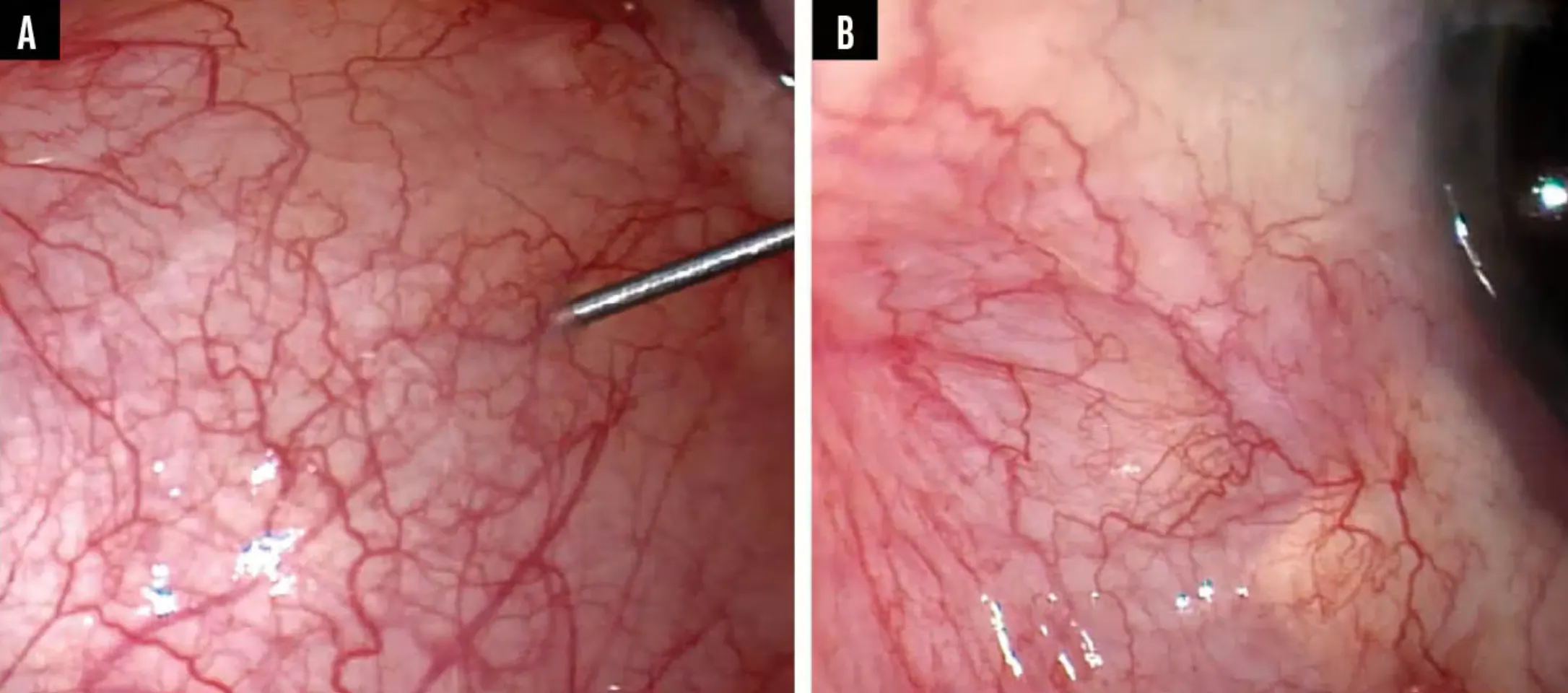
Figure 2. Before (A) and after (B) decompression of a large bleb that was causing restrictive strabismus.
TUBE SWEEP
Sometimes, a beautifully positioned tube becomes occluded by the iris postoperatively. The sweeping technique involves both a needle and a laser but is otherwise straightforward. A 30-gauge needle is passed into the AC and then used to gently sweep the iris away from the tube opening until the lumen is unobstructed. Function often resumes immediately, but the iris may eventually reocclude the tube.
To reduce the likelihood of recurrence, a focal argon laser iridoplasty may be performed adjacent to the tube using settings similar to those employed for plateau iris syndrome: approximately 200 mW of power with a spot size of 150 to 200 µm. The goal is to contract and stiffen the iris tissue surrounding the tube, thus reducing its tendency to prolapse back into the opening.
HYPHEMA DRAINAGE
It is not uncommon to encounter a patient with a hyphema after MIGS or a patient with traumatic hyphema and elevated IOP who would traditionally be considered for an AC washout.7 If the IOP is very elevated, such as 50 mm Hg or higher, one should almost reflexively attempt drainage because time is of the essence.
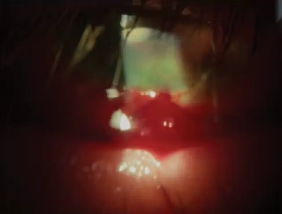
My (I.O.) technique was inspired by a video from the CataractCoach series from Uday Devgan, MD, featuring an innovative slit-lamp procedure developed by Ramesh Ayyala, MD.8 A microvitreoretinal blade is used to create an inferior paracentesis at the slit lamp. Through this paracentesis, a 30-gauge needle is introduced, and a large air bubble is injected into the AC. Frequently, the hyphema immediately begins to egress through the inferior wound (Figure 3). If additional drainage is required, the wound may be gently burped, or additional air may be injected. A medium-sized air bubble is left in the eye to repressurize the AC and prevent further bleeding.
If the blood has not clotted excessively, a substantial portion of the hyphema can often be evacuated. This approach can avoid multiple trips to the OR, and patients typically experience an improvement in their visual acuity once the bubble dissipates. If active bleeding persists, the procedure can be repeated as necessary.
CONCLUSION
One of the strengths of glaucoma surgery is its creativity and adaptability. Sometimes, the most valuable intervention is the one that the surgeon can perform safely and effectively right at the slit lamp.
1. Childers CP, Maggard-Gibbons M. Understanding costs of care in the operating room. JAMA Surg. 2018;153(4):e176233. doi:10.1001/jamasurg.2017.6233
2. Hosler MR, Scott IU, Kunselman AR, Wolford KR, Oltra EZ, Murray WB. Impact of resident participation in cataract surgery on operative time and cost. Ophthalmology. 2012;119(1):95-98. doi:10.1016/j.ophtha.2011.06.026
3. Sit AJ, McLaren JW. Measurement of episcleral venous pressure. Exp Eye Res. 2011;93(3):291-298. doi:10.1016/j.exer.2011.05.003
4. Mardelli PG, Lederer CM, Murray PL, Pastor SA, Hassanein KM. Slit-lamp needle revision of failed filtering blebs using mitomycin C. Ophthalmology. 1996;103(11):1946-1955. doi:10.1016/s0161-6420(96)30403-x
5. Tang M, Gill N, Tanna AP. Incidence and outcomes of hypertensive phase after glaucoma drainage device surgery. Ophthalmol Glaucoma. 2024;7(4):345-351. doi:10.1016/j.ogla.2024.03.006
6. Molteno AC, Fucik M, Dempster AG, Bevin TH. Otago Glaucoma Surgery Outcome Study: factors controlling capsule fibrosis around Molteno implants with histopathological correlation. Ophthalmology. 2003;110(11):2198-206. doi:10.1016/S0161-6420(03)00803-0
7. Walton W, Von Hagen S, Grigorian R, Zarbin M. Management of traumatic hyphema. Surv Ophthalmol. 2002;47(4):297-334. doi:10.1016/s0039-6257(02)00317-x
8. Devgan U. CataractCoach 2865: how to drain hyphemas at the slit lamp microscope. 2026. Accessed September 8, 2026. www.youtube.com/watch?v=r6jqtbt-9Es