Key Takeaways
- Zonular instability can lead to the dislocation of primary and secondary IOLs and raises concern about glaucomatous progression.
- The potential consequences of a malpositioned IOL, such as disease progression and chronic inflammation, rather than the dislocated lens itself are key factors in the decision whether to intervene surgically.
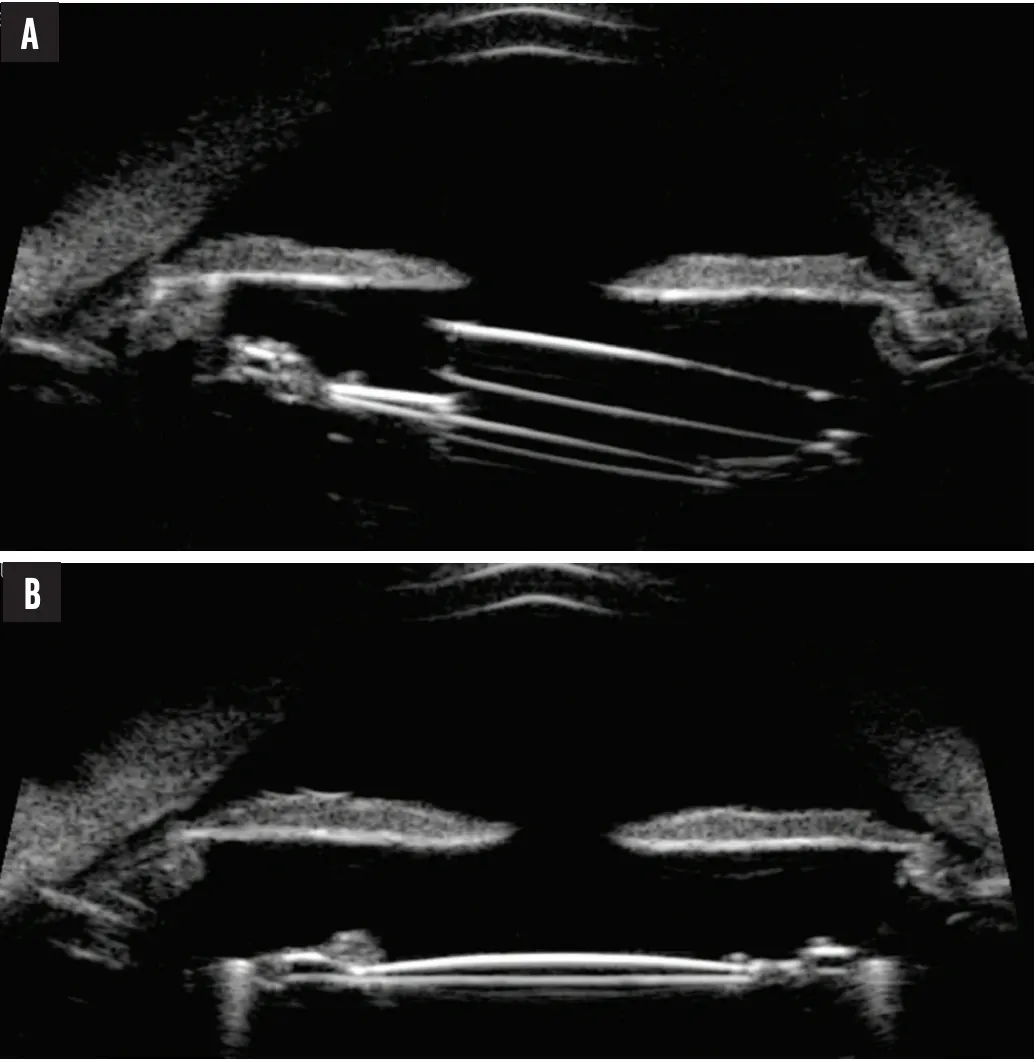
Figure 1. UBM shows a tilted and subluxated piggyback IOL in the sulcus of the right eye (A) and a posteriorly displaced IOL–capsular bag complex in the left eye (B).
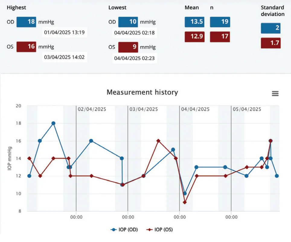
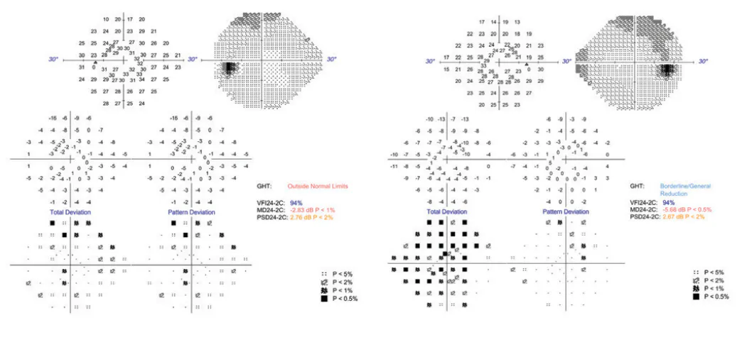
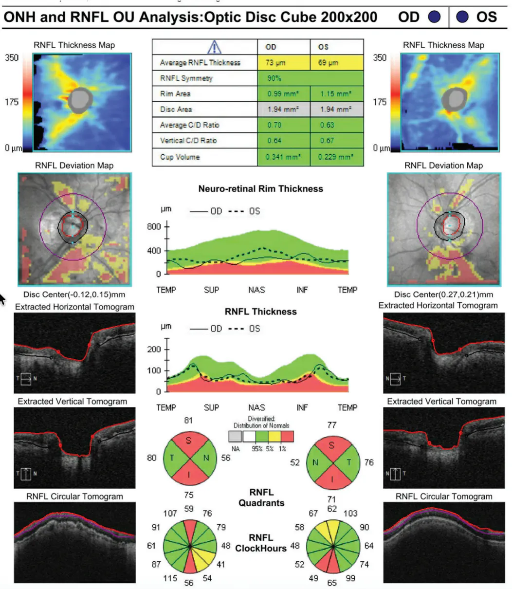
Figure 4. Visual field testing shows no specific loss in the right eye and possible early superior glaucomatous changes in the left eye.
SAHAR BEDROOD, MD, PHD
The bilateral zonular instability raises suspicion for underlying pseudoexfoliation syndrome, which will likely worsen over time.
In the right eye, zonular weakness has resulted in tilt and subluxation of both the piggyback IOL and the capsular bag–IOL complex. Chronic iris chafing has caused pigment dispersion, IOP fluctuation, iris transillumination defects, and early RNFL loss, consistent with mechanical pigmentary glaucoma. Without intervention, ongoing pigment release and IOP instability would increase the patient’s risk of irreversible glaucomatous vision loss.
In the left eye, significant zonular instability has caused subluxation of the entire IOL–capsular bag complex. The optic bisects the visual axis, leading to a symptomatic visual disturbance. Progressive destabilization would likely result in complete dislocation of the lens, necessitating urgent removal with vitreoretinal assistance.
Proactive surgical intervention is warranted in both eyes.
Updated biometry measurements for the right eye would be obtained. Both the piggyback IOL and the capsular bag–posterior chamber IOL (PCIOL) complex would be removed, and an appropriately powered IOL would be implanted. Either sutureless intrascleral haptic fixation (ISHF) of a three-piece lens (SofPort LI61AO, Bausch + Lomb) using the Yamane technique or transscleral fixation of a one-piece lens such as an enVista (model MX60, Bausch + Lomb) with PTFE or polypropylene sutures would be performed.
If the IOL in the left eye is well centered in the bag, transscleral fixation of the entire lens–capsular bag complex might preserve the current implant and restore stability without the need for more extensive removal of the entire lens.
Preoperatively, the patient would be counseled that her postoperative recovery could be complex. Potential complications include cystoid macular edema, IOL tilt, vitreous hemorrhage, retinal detachment, and IOP spikes or fluctuations that might ultimately require glaucoma drainage device (GDD) implantation. Lifetime monitoring for lens stability and IOP fluctuations would be warranted.
LEON W. HERNDON JR, MD, AND YANNEK LEIDERMAN, MD, PHD
The patient has a history of pigment dispersion, but fortunately, no significant glaucomatous atrophy is evident. A description of the optic nerves would facilitate a decision on glaucoma surgical intervention, but the RNFL and visual field studies are reassuring. She has experienced bilateral IOL subluxation, and her concern is refractive in nature.
The left eye is more straightforward in that displacement of the IOL-CTR complex is optically significant. Examination findings consistent with uveitis-glaucoma-hyphema syndrome are lacking, and visual field testing is inconclusive for glaucoma in the setting of RNFL thinning. It would therefore be reasonable to intervene surgically with the goal of addressing the refractive component while minimizing the potential for glaucomatous progression associated with IOL placement. The removal of a subluxated IOL-CTR complex carries a risk of vitreous loss. Our preference would be to combine a pars plana vitrectomy with removal of the IOL-CTR complex and implantation of a three-piece IOL via tunnel-based ISHF. Although sutureless scleral fixation techniques such as those described by Yamane and colleagues have surged in popularity, we prefer ISHF to minimize the risks of haptic exposure and endophthalmitis.
The right eye exhibits subluxation of the piggyback IOL. The presence of iris transillumination defects in conjunction with asymmetric pigmented cells in the anterior chamber is highly suggestive of an IOL-induced mechanical component that is exacerbating the patient’s inherent pigment dispersion. Alleviating the IOL-induced chafing might decrease the risk of glaucomatous progression. An IOL exchange could be performed if the capsular-zonular diaphragm is adequate, but the dislocated sulcus IOL might increase the risk of primary IOL dislocation postoperatively. If there is any evidence of zonular instability, both the sulcus IOL and one-piece IOL would be explanted, and sutureless scleral fixation of a three-piece IOL would be performed via a tunnel-based approach. Because the eye may require a GDD in the future, flange-based and scleral-sutured haptic fixation techniques would be avoided.
JOSEPH F. PANARELLI, MD
To my mind, the patient’s age, glaucoma severity, and endothelial cell count are the most important considerations. A younger patient might achieve the greatest benefit from a surgically intense procedure that resolves the problem and achieves stability for decades to come, whereas a quick fix might be more suitable for a patient older than this one.
The IOP in each of the patient’s eyes has been relatively stable, and she retains peripheral vision. OCT appears to show a nasal artifact in each eye caused by shifting peaks, as evident on the RNFL thickness plot. Despite the pigment dispersion and findings akin to uveitis-glaucoma-hyphema syndrome in the right eye, neither is an urgent concern. Deciding how to proceed could be challenging because the outflow facility may already be damaged.
At this point, I would do nothing to address the IOP in either eye. It could be argued, however, that a prophylactic goniotomy or the implantation of a GDD in either or both eyes might be of benefit.
Given the disease stability demonstrated to date and the complexity of IOL surgery, the patient would be closely monitored and counseled preoperatively on her potential need for additional future surgery.
The endothelial cell counts look good, so there are numerous options for IOL fixation. The decision largely depends on the patient’s preferences and ability to function in the postoperative period. Is her top priority the quickest possible postoperative recovery, or would she prefer to undergo more extensive surgery to minimize the risk of a return trip to the OR?
The entire IOL–capsular bag complex in the left eye could be fixated to the iris. This strategy would likely achieve the fastest postoperative visual recovery with the lowest amount of risk. It would also allow the more complex right eye to be treated sooner than would other fixation strategies.
My preference for the right eye would be to explant the piggyback IOL; remove the PCIOL-CTR complex, which appears on UBM to be displaced; and perform scleral fixation of a new IOL. I would also consider removing the IOL in each eye and leaving the patient aphakic (correcting her ametropia with contact lenses). Although the latter approach is not a popular strategy, it would minimize the risk of complications because these eyes are prone to bleeding and other complications that require additional surgery. It is important, however, not to base surgical decisions on the one case that did not go as planned. A reasonable, individualized assessment of risks and benefits must be conducted.
PRADEEP RAMULU, MD, PHD
The patient’s visual symptoms are likely driven by IOL-related issues; most individuals do not notice visual field damage until the visual field index drops into the 80s, whereas a tilted piggyback IOL and displaced PCIOL are typically visually symptomatic.
The patient’s visual field tests and OCT scans may indicate early glaucoma, but they may also reflect axial myopia. Supporting this idea are the temporal displacement of the superior and inferior RNFL bundles, the nonspecific pattern of visual field loss in the right eye and the diffuse visual field loss in the left eye (both seen with myopia), and the reassuring results of home tonometry. It remains possible, however, that the patient experienced elevated IOP years ago that resolved as accommodation was lost or as a result of cataract surgery.
Given her stable unmedicated IOP, I would not recommend glaucoma surgery at this time. Although some have postulated an increased risk of glaucoma with vitrectomy, eyes undergoing a pars plana vitrectomy for simple conditions such as an epiretinal membrane do not appear to be at higher risk of developing glaucoma than contralateral eyes.1
I would address the left eye first. The IOL would be explanted, and scleral fixation of a new IOL with a partial or full vitrectomy would be performed. The best approach to the right eye depends on the patient’s desire for spectacle independence. If she is willing to wear glasses or contact lenses, removal of the piggyback IOL should be adequate. If the surgical and refractive outcomes in the left eye are good, she could also consider the removal of both the PCIOL and piggyback IOL followed by scleral fixation of a new IOL.
WHAT I DID: DEVESH K. VARMA, MD, FRCSC
Despite the evidence of pigment dispersion and iris chafing in the right eye, the clinical findings had remained stable over time, and there was no compelling evidence of ongoing damage. Given the potential risks associated with surgery, continued surveillance was deemed the most appropriate course of action.
For the left eye, surgical management was recommended because the risk of future deterioration appeared to outweigh the risks of intervention. The initial plan was to reposition the existing lens but to modify the approach if necessary based on intraoperative findings. During surgery, the degree of instability proved greater than anticipated. The IOL–capsular bag complex was therefore removed, an anterior vitrectomy and peripheral iridotomy were performed, and an Artisan IOL (Ophtec) was implanted. The postoperative course was uncomplicated aside from a transient IOP elevation that responded to treatment. The patient was pleased with her visual outcome, particularly her near vision.
As this case demonstrates, the indication for surgical intervention is not the presence of an abnormally located IOL or a dislocated IOL but the likelihood that the abnormality will result in vision loss, glaucomatous progression, inflammation, or ongoing instability. When multiple pathologies coexist, the challenge is determining what should be fixed rather than what can be fixed.
1. Lalezary M, Kim SJ, Jiramongkolchai K, Recchia FM, Agarwal A, Sternberg P Jr. Long-term trends in intraocular pressure after pars plana vitrectomy. Retina. 2011;31(4):679-685. doi:10.1097/IAE.0b013e3181ff0d5a