The Optic Nerve Analysis software for Optovue’s spectral-domain optical coherence tomography (SD-OCT) devices provides clinicians with three sets of data for glaucoma evaluation. The optic nerve head (ONH) analysis measures the disc area, the rim area, and the cup-to-disc ratio. The peripapillary retinal nerve fiber layer (RNFL) analysis measures the average RNFL thickness, the hemifield RNFL thickness, and the quadrant RNFL thickness. The macular region ganglion cell complex (GCC) analysis measures the average GCC thickness, the hemifield GCC thickness, the focal loss volume, and the global loss volume. The ONH and RNFL parameters are derived from the ONH scan, and the GCC parameters are derived from the GCC scan (Figure).
The measurement parameters from these three sets of analysis are automatically compared to the OCT’s normative limits, and the results are color-coded for “within normal limits” (green), “borderline” (yellow), and “outside normal limits” (red). The normative limits are always adjusted for age and, in the cases of ONH and RNFL parameters, are also adjusted for optic disc size (Figure).
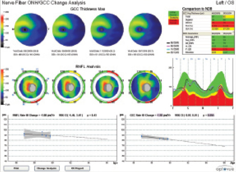
Figure. The RNFL/ONH and GCC report with change analysis.
The Optic Nerve Analysis with Optovue’s SD-OCT devices is repeatable and reproducible, with the coefficient of variation not exceeding 2.1% for the average RNFL thickness and not exceeding 1.7% for the average GCC thickness in healthy and glaucomatous eyes (data on file with Optovue). Trend analysis to estimate the rates of change of the RNFL and the GCC is also provided with the company’s Avanti and iVue devices for longitudinal assessment of the optic nerve.
Jason Bacharach, MD, is the director of research at North Bay Eye Associates in Sonoma, California, and vice chair of the Glaucoma Department at California Pacific Medical Center in San Francisco. He acknowledged no financial interest in the products or companies mentioned herein. Dr. Bacharach may be reached at (707) 762- 6622; jb@northbayeye.com.