Key Takeaways
- Whales, squid, and deep-sea fish illustrate a central principle: Ocular survival under pressure depends on structure.
- The future of glaucoma therapy may require ophthalmologists not only to lower pressure but also to better understand, and perhaps one day modify, the tissues that determine how that pressure is experienced.
- Research is being conducted to explore whether altering the biomechanical environment of the optic nerve head could alter its susceptibility to IOP-related injury.
A few years ago, while I (D.F.) was watching The Wild Kratts children’s show with my daughter, I learned that sperm whales routinely dive thousands of feet below the ocean surface and hold their breath for more than an hour while hunting giant squid in near darkness. While my daughter focused on the on-screen battle between whale and squid, I found myself wondering: How does a mammalian eye tolerate such an extreme environment without developing glaucoma?
At depths approaching 6,000 feet, external hydrostatic pressure is immense. The lesson, however, is not that whales experience IOP in the way humans with glaucoma do. Rather, the eye is a biomechanical organ, and nature has solved the problem of pressure tolerance in multiple ways.
DIFFERENCES IN THE CORNEA AND SCLERA
Whale eyes can withstand extreme conditions in part because of differences in the cornea and sclera. Cetacean corneas and sclerae are substantially thicker than those of humans (Figure). The whale cornea shares broadly similar collagen composition, whereas the whale sclera is denser and more robust.1 In some species of whales, the posterior sclera is extraordinarily thick, forming a reinforced outer coating that helps preserve globe integrity. The whale sclera is not merely a passive container but a pressure-resistant biomechanical scaffold.
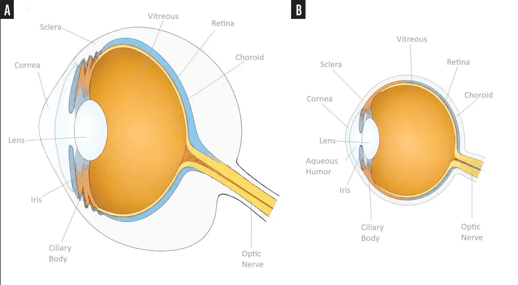
Figure. A comparison of whale eye anatomy (A) and human eye anatomy (B). Whales have the largest eyes among mammals, with an axial length of around 9 to 13 cm, whereas average human eyes have an axial length of around 23 to 24 mm. A human cornea is about 500 µm thick, whereas a whale cornea is about 2 to 3 mm thick. The sclera is proportionally much thicker in whale eyes; it measures about 4 to 5 cm, approximately 25% to 33% of the diameter of the eye.
EVOLVED STRUCTURAL STRATEGIES
An examination of nonmammalian marine species adds another layer of intrigue. Deep-sea fish and cephalopods are exposed to similarly extraordinary hydrostatic pressures but have evolved distinct strategies to preserve neural, vascular, and optical function. Squid, for example, developed camera-like eyes through convergent evolution to arrive at a superficially similar optical design through entirely different developmental pathways. Their enormous eyes are optimized for deep-sea light detection, including faint bioluminescent cues. Their supporting tissue is not collagenous sclera but a cartilaginous scleral support system derived from distinct tissues. Their relevance to human glaucoma is therefore less direct. Rather than model mammalian optic nerve biomechanics, these species show that ocular stability under pressure can be achieved through very different structural strategies.
INSIGHTS INTO HUMAN GLAUCOMA
The broader lesson for glaucoma specialists is not that whales, squid, and fish are interchangeable models. They are not. Rather, they remind us that pressure tolerance is a property of tissue architecture. Across species, ocular shape, neural integrity, and visual function are preserved through coordinated structural adaptations. For the management of human glaucoma, this perspective encourages us to look beyond IOP alone and ask why some optic nerves tolerate pressure whereas others deform and whether the sclera, lamina cribrosa, and peripapillary connective tissues are as important as the pressure itself.
It has become increasingly clear that scleral biomechanics play an important role in glaucoma. The peripapillary sclera provides mechanical support to the optic nerve head, and the thickness, stiffness, collagen organization, and remodeling capacity of the former influence how IOP-related forces are transmitted to the lamina cribrosa and retinal ganglion cell axons. Studies have reported differences in collagen organization and scleral remodeling in glaucomatous eyes, although it remains unclear whether these are predisposing factors, adaptive responses, or both.2
The sclera is not a static shell. Its stiffness and deformation characteristics change in response to IOP. As IOP rises, the sclera deforms and transmits mechanical forces to the optic nerve head. Work using animal models, 3D histomorphometry, and OCT-based imaging suggests that lateral scleral deformation may exert a greater biomechanical effect on the lamina cribrosa than direct posterior displacement from IOP alone.2 This distinction matters. Glaucoma is often conceptualized as pressure pushing the optic nerve backward. A more complete model is pressure-induced deformation of a complex connective tissue system, with the sclera stretching and redistributing forces around the optic nerve head. In donor eye studies, thinner or more compliant posterior sclerae are more deformed by equivalent pressure loads, producing greater mechanical strain at the optic nerve head.3 This has been proposed as one mechanism underlying normal-tension glaucoma and increased susceptibility to glaucoma due to connective tissue disorders.
Researchers have begun targeting scleral and laminar biomechanics as potential therapeutic pathways, but early results have been mixed. In some models, modifying scleral stiffness produced different effects at low versus high pressures.4 Other studies attempting to strengthen the posterior sclera through crosslinking have produced paradoxical findings, including increased susceptibility to retinal ganglion cell damage.5 These results warrant caution.
A whale sclera is not simply a stiffer human sclera but part of an integrated system shaped by evolution. That may be the most important lesson from comparative anatomy. Evolution rarely solves a problem by changing a single variable. The whale eye, squid eye, and deep-sea fish eye represent coordinated solutions to environmental pressure. Some emphasize resistance to deformation, others prioritize light capture, and others modify retinal structure, ocular geometry, or vascular support.
For glaucoma, this broader perspective is useful. We should continue to lower IOP because it is the only proven modifiable risk factor, but we should also think beyond IOP alone. Why does one patient’s disease progress at 14 mm Hg, whereas another’s remains stable at 24 mm Hg? Why do some optic nerves tolerate pressure but others deform? Why do some eyes show rapid functional loss despite modest IOP? Part of the answer may lie in the biomechanical behavior of the sclera, lamina cribrosa, and peripapillary connective tissues.
CONCLUSION
I (D.F.) do not usually look to children’s television for research inspiration, but that afternoon watching The Wild Kratts with my daughter reminded me that some of the most interesting questions in medicine begin with observations from the natural world. Nature has run pressure experiments for millions of years. Whales, squid, and deep-sea fish illustrate a central principle: Ocular survival under pressure depends on structure.
The future of glaucoma therapy may require us not only to lower pressure but also to better understand, and perhaps one day modify, the tissues that determine how that pressure is experienced. This concept is already shaping work in our group as we explore whether altering the biomechanical environment of the optic nerve head could alter its susceptibility to IOP-related injury.
From whales to humans, the next wave of glaucoma research may come not only from looking inside the eye but at the shell that holds it together.
Authors’ AI disclosure: An AI language model (GPT-5.5, OpenAI) was used to assist in the creation of the figure in this manuscript. The authors sketched and labeled the figure and used AI to refine the quality of the illustration.
1. Vecino E, Ruzafa N, Pereiro X, Zulueta A, Sarmiento A, Díez A. The extracellular matrix of the human and whale cornea and sclera: implications in glaucoma and other pathologies. In: Madhurapantula RS, Orgel J, Loewy ZG, eds. Extracellular Matrix: Developments and Therapeutics. IntechOpen; 2021. doi:10.5772/intechopen.97023
2. Jia X, Yu J, Liao SH, Duan XC. Biomechanics of the sclera and effects on intraocular pressure. Int J Ophthalmol. 2016;9(12):1824-1831. doi:10.18240/ijo.2016.12.21
3. Coudrillier B, Tian J, Alexander S, Myers KM, Quigley HA, Nguyen TD. Biomechanics of the human posterior sclera: age- and glaucoma-related changes measured using inflation testing. Invest Ophthalmol Vis Sci. 2012;53(4):1714-1728. doi:10.1167/iovs.11-8009
4. Murienne BJ, Jefferys JL, Quigley HA, Nguyen TD. The effects of glycosaminoglycan degradation on the mechanical behavior of the posterior porcine sclera. Acta Biomater. 2015;12:195-206. doi:10.1016/j.actbio.2014.10.033
5. Kimball EC, Nguyen C, Steinhart MR, et al. Experimental scleral cross-linking increases glaucoma damage in a mouse model. Exp Eye Res. 2014;128:129-140. doi:10.1016/j.exer.2014.08.016