


Key Takeaways
- Given the declining rates of trabeculectomy cases worldwide, learning the nuances of this surgery has become more challenging.
- With a systematic approach and appropriate experience, trabeculectomy remains a reproducible and effective surgery.
- Attention to small cues and technical details guides the surgeon through the steepest parts of the learning curve.
Trabeculectomy remains the benchmark against which every glaucoma filtration procedure is measured. Even as minimally invasive options multiply, guarded filtration surgery continues to achieve some of the lowest IOPs of any current glaucoma procedure and remains an indispensable option for advanced and refractory disease.1,2 Meticulous technique can help to lower the risk of complications associated with this surgical approach.3
With the widespread adoption of MIGS, however, ophthalmology trainees may be performing fewer trabeculectomies.4,5 In an 11-year period, US glaucoma fellows experienced a 55.9% decline in trabeculectomy exposure, coinciding with a 128.6% rise in MIGS procedures.5 This pattern is also evident at our center, King Khaled Eye Specialist Hospital & Research Center (KKESH), where total trabeculectomy volume decreased from 116 cases performed in 2019 to 47 cases performed in 2024 (Figure 1).
Importantly, trabeculectomy can be performed in virtually any surgical setting, making it an essential technique for trainees to master and a particularly valuable option in resource-limited environments. This article distills practical, step-by-step tips for trabeculectomy with mitomycin C (MMC).
PATIENT SELECTION
Careful patient selection is an important determinant of trabeculectomy outcomes. Beyond the standard clinical indications such as inadequate IOP control on maximum tolerated medical therapy, progressive optic nerve damage, and visual field loss, patient-related factors must be considered. Our preference is not to offer trabeculectomy to patients who are unable to attend weekly follow-up visits in the first 3 postoperative weeks, because this period is critical for IOP titration, suture management, and the early detection of complications, such as hypotony and bleb failure.
Patients who have difficulty adhering to prescribed medical therapy may also struggle to comply with a postoperative topical steroid regimen, which is vital for trabeculectomy success. For such patients, alternative surgical approaches or a structured period of supervised medical therapy before scheduling surgery merit consideration.
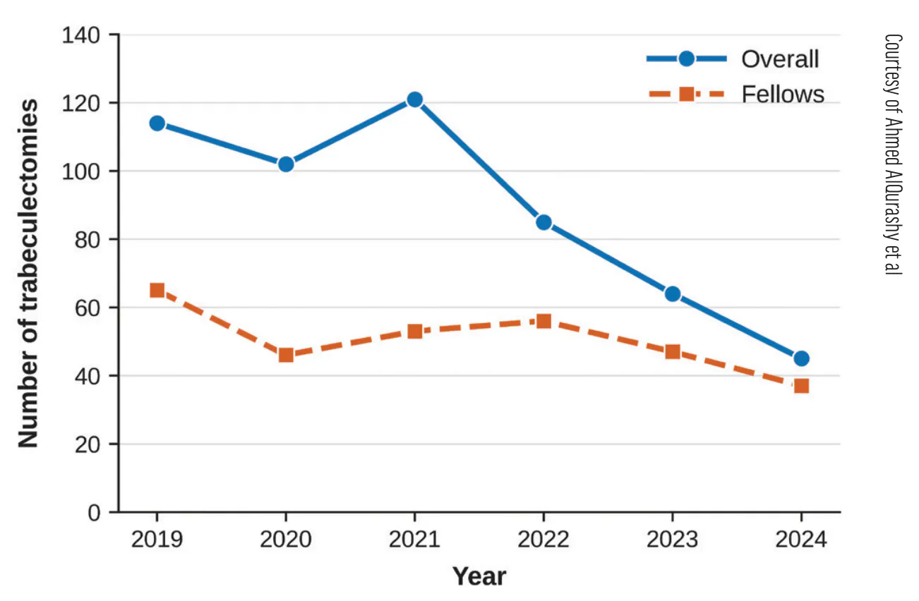
Figure 1. Annual trends of trabeculectomy procedures at KKESH, showing a gradual decline from 2019 to 2024 among surgeons and glaucoma fellows (unpublished data).
SURGICAL TECHNIQUE
Pupillary Constriction and Site Selection
One drop of pilocarpine 2% is instilled preoperatively. A miotic pupil draws the iris taut, protects the lens, and can ultimately make the peripheral iridectomy cleaner. A superior surgical site near the 12 clock position is acceptable in older patients, who may be more likely to receive a glaucoma drainage device as a subsequent surgery rather than to undergo another filtering surgery. In children and younger patients, we prefer a superonasal approach. Although nasal access can be slightly more difficult, this approach preserves the superotemporal conjunctiva for possible subsequent filtering procedures.
Exposure and Traction Suture
Adequate exposure of the superior conjunctiva is key. Our preference is to use a Lieberman adjustable speculum with open K-wire blades (Keeler). In rare circumstances, if the speculum sits tightly or compromises exposure of the superior conjunctiva, a Jaffe wire lid retractor can be used, or even a 4-0 silk lid traction suture may be passed directly through the grey line. A 7-0 polyglactin clear corneal traction suture (Vicryl, Ethicon) is then placed superiorly. The traction suture is set slightly more anteriorly than usual to keep it clear of the scleral flap and the corneal entry sites that could later be used for releasable sutures (discussed later under Preplacement of Scleral Flap Sutures).
Five Tips for Trabeculectomy
- 1. Minimize unnecessary manipulation of the conjunctiva, especially in eyes with thin tissues.
- 2. Aim for a good posterior dissection with a broad application of mitomycin C.
- 3. Use an air bubble (through a paracentesis) to maintain the anterior chamber during the sclerostomy.
- 4. Aim for no flow (or minimal flow on posterior pressure) through the flap after securing the sutures.
- 5. Use a method of watertight conjunctival closure.
Conjunctival Peritomy
The intended surgical field is marked superiorly over approximately 2 to 3 clock hours. A fornix-based peritomy can be fashioned against the limbus or 1 mm behind the limbus, leaving a small strut of conjunctiva for ease of closure. Our preference is the former approach. The first cut in the conjunctiva is made horizontally with Vannas scissors, parallel to the limbus. When exposure is adequate, an incision at the limbus eliminates the need for a relieving radial incision and minimizes the risk of leaks through radial incisions. Subconjunctival injection of 0.5% bupivacaine at the site of the first cut helps separate the Tenon capsule from the sclera, indicates conjunctival mobility, and provides some postoperative analgesia, which is especially useful in children.
Sharp Westcott scissors are then used to dissect along the limbus. An extensive subconjunctival dissection is performed with blunt Westcott scissors to aim for a diffuse posterior bleb. During this step, the scissor blades can be oriented downward so that they follow the curve of the sclera. During dissection, it is better to hold the Tenon capsule rather than the conjunctival tissue to avoid conjunctival tears (a common mistake among inexperienced trainees). In patients with thin conjunctiva, a Weck-Cel sponge (BVI) in the nondominant hand can be used to retract the conjunctiva, and/or nontoothed forceps can be used to hold the conjunctiva when needed. Care should be taken to avoid dissection directly over the superior rectus muscle, whose insertion lies approximately 7.7 mm behind the limbus, because trauma to the associated vasculature can cause bleeding.
MMC Application
Our preference is to use a single dry surgical sponge that is grasped with serrated forceps along its full length to prevent it from slipping during insertion. The sponge is introduced from the corneal side and slid as far posteriorly as possible under the Tenon layer. MMC at a concentration of 0.2 mg/mL or 0.4 mg/mL is injected through a cannula underneath the sponge and left for 3 minutes. The injection of MMC without a sponge is a reasonable approach and may produce a more diffuse, less vascularized bleb with comparable success rates.6 We prefer to use a large sponge to ensure that the conjunctival dissection is adequate (with enough space created) and to prevent MMC from spilling over the conjunctiva and wound edges. Use of a single sponge, rather than multiple small ones, facilitates removal (small sponges can occasionally be difficult to retrieve from the superior conjunctival fornix). The concentration of MMC is usually individualized according to risk factors for failure. A higher concentration (0.4 mg/mL) may be more appropriate in younger patients and those with previous failed filtration surgery, whereas a lower concentration (0.2 mg/mL) is generally suitable for eyes with thin conjunctiva and a lower scarring response (typically older patients). After the sponge has been removed, the area is irrigated copiously with balanced salt solution. Contact between the MMC and the conjunctival wound edge is avoided to reduce the risk of a leak, and the edges can be dried with a Weck-Cel sponge.
Hemostasis
The MMC dwell time is used efficiently for performing hemostasis, with attention focused on the prospective flap edges. A wet-field bipolar cautery (Wet-Field Hemostatic Eraser Bipolar, BVI) with an eraser tip is used at a power setting of 15% to 30%. Cautery is kept to a minimum because excessive cautery can cause tissue contraction and scleral thinning that may complicate flap dissection. A 3-mm Tooke corneal knife (Katena) can be used to clear residual Tenon adhesions from the scleral surface and create a clean bed for the trabeculectomy flap.
Superficial Scleral Flap
A rectangular flap measuring approximately 4 to 5 mm wide (parallel to the limbus) and 2 to 2.5 mm long is marked. There are two approaches to creating the flap: (1) under direct visualization using a Beaver 6600 Mini-Blade (BVI) or similar blade or (2) with less direct visualization using a crescent blade under the sclera. We prefer the latter approach because we have found it creates a predictable, homogeneous flap. In addition, if bleeding occurs, it does not hinder the dissection (Figure 2).
The posterior horizontal edge of the flap is incised first with a SupraSharp blade (Surgistar) while the tissue is stabilized with 0.12-mm forceps. The depth is checked, and the goal is one-third to one-half of the scleral thickness. A crescent blade is then used to raise the flap. The heel of the blade is kept down so that the blade remains faintly visible through the tissue, confirming a partial-thickness plane. The crescent blade is advanced anteriorly with a fine microcircular painting motion, carried to the limbus, and then extended gently to the sides until it reaches the left and right flap edges. When it is moving laterally, the crescent blade should not be tilted but instead kept flat to preserve a single uniform plane. The more anteriorly the dissection proceeds, the farther the heel is dropped to avoid early perforation into the anterior chamber. The flap margins are then created with a SupraSharp blade and 0.12-mm forceps, allowing the flap to fold forward cleanly and reveal the limbus. For this dissection, small sideways cuts can be made (from the inside outward) while the flap is lifted gently.
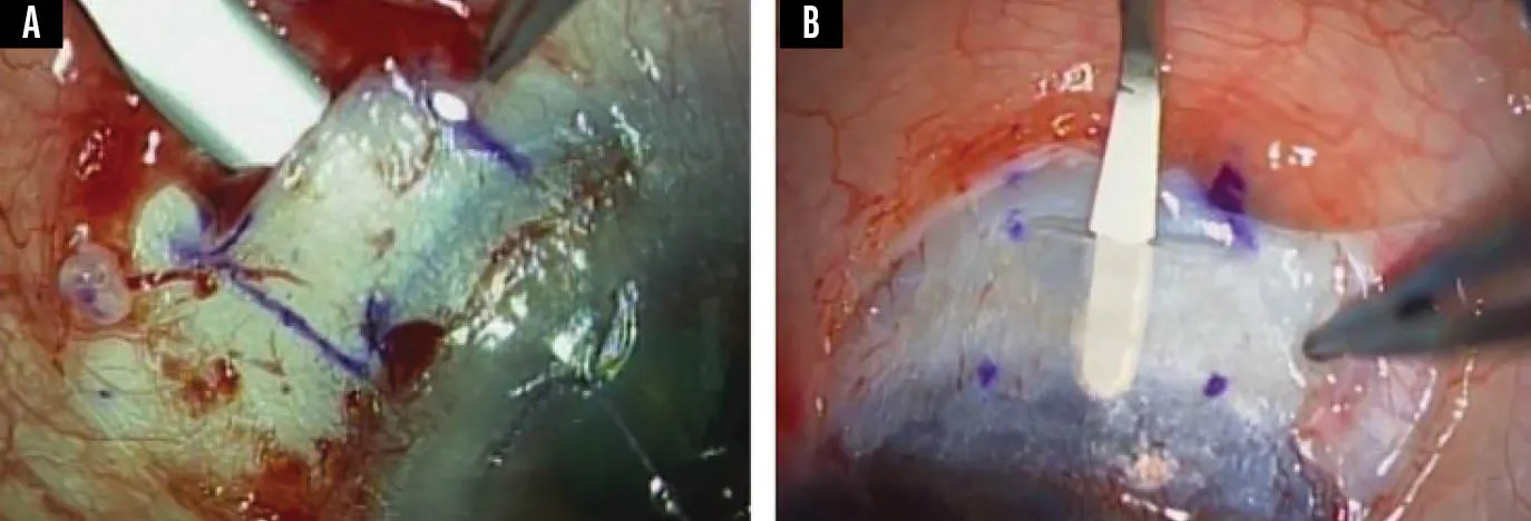
Figure 2. Dissection of the scleral flap with a crescent blade. Bleeding from the episcleral vessels does not hinder the dissection (A). Use of the same technique in a child’s eye with thin sclera (B).
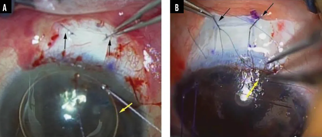
Figure 3. Methods of scleral flap closure. Fixed sutures (black arrows) require argon laser suture lysis postoperatively (A). Note the air bubble (yellow arrow) maintaining the AC. Releasable sutures can be removed at the slit lamp and do not require lysis with a laser (black arrows, B). The proximal part of the suture (yellow arrow) is placed within a groove for subsequent removal at the slit lamp.
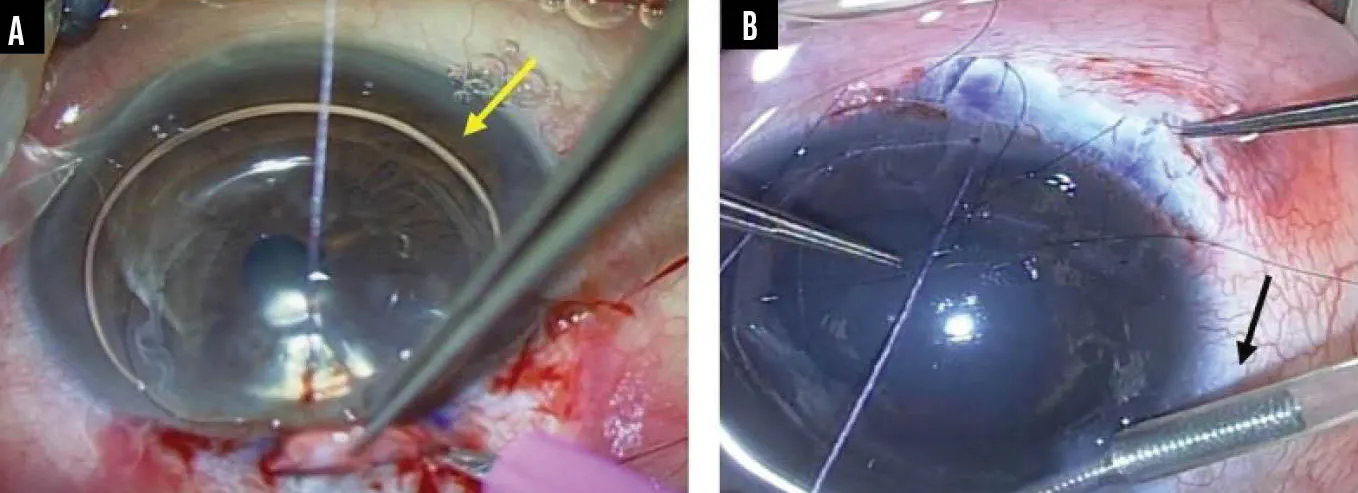
Figure 4. Maintaining the AC during trabeculectomy. The KKESH air bubble technique involves placing an air bubble (yellow arrow) in the AC through a paracentesis before performing the sclerostomy (A). Use of an AC maintainer (black arrow) in a child’s buphthalmic eye (B).
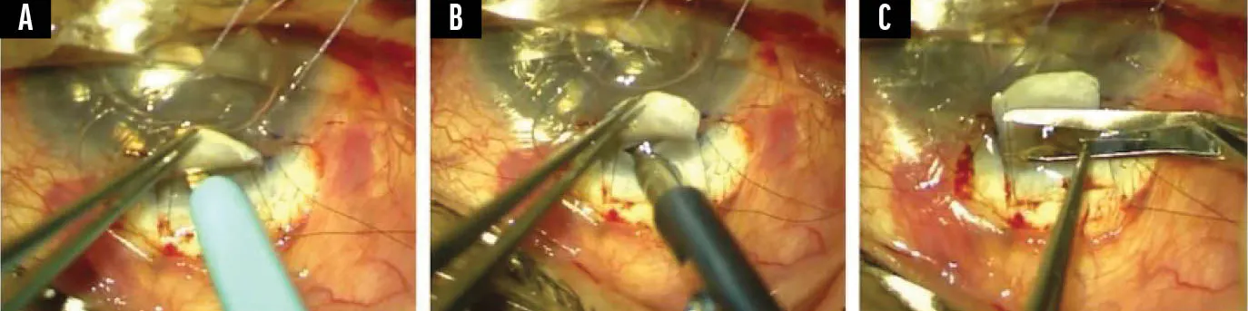
Figure 5. Performing the sclerostomy. A wide scleral incision facilitates insertion of the punch (A). The Kelly punch should hook the scleral ledge and be as vertical as possible before a punch is taken (B). The far peripheral iris is grasped, and a single clean cut is made at the base of the iris to create a sufficient, clean triangular iridectomy (C).
Preplacement of Scleral Flap Sutures
Although both fixed and releasable sutures can be used for trabeculectomy (Figure 3), the latter offer some advantages. Postoperative lysis with an argon laser is not required, making releasable sutures useful in children and patients who cannot look down easily. Releasable sutures also facilitate postoperative manipulation in settings where a laser is not available. Two scleral flap sutures (fixed or releasable) are preplaced before the anterior chamber (AC) is entered, while the field is dry and the globe is firm.
Paracentesis and Preparation for Entry
A temporal paracentesis is created, and the AC is filled with air (rather than balanced salt solution) to keep the AC formed and prevent its collapse during sclerostomy creation (the KKESH air bubble technique; Figure 4A). If the sclerostomy is made gently, without excessive pressure on the eye, the bubble should maintain the AC through the sclerostomy and iridectomy. In bupthalmic or very soft eyes, an AC maintainer may be used instead (Figure 4B).
The traction suture is then relaxed slightly. Before the eye is entered, the Kelly punch and de Wecker scissors are confirmed to be working to prevent fumbling with a faulty instrument inside an open eye. The sclerostomy should be made efficiently, minimizing how long the eye is in a hypotonous state.
Anterior Chamber Entry
An approximately 3-mm entry into the AC is made at the anterior margin of the flap bed into clear cornea with the SupraSharp blade held vertically (or at least obliquely) (Figure 5A). An adequate incision facilitates insertion of the punch. If the entry site is too shallow, the punch will be difficult to insert. A controlled, well-oriented sclerostomy sets up a clean block excision.
Block Excision
The trabecular block is removed with a Kelly punch. Our preference is to use a 0.70- to 0.75-mm micro-Kelly punch (eg, Rumex microtrabeculectomy punch, Titan Medical, Katena). The instrument is introduced obliquely and then positioned as vertically as possible before a punch is taken; one or two bites are generally sufficient to create an adequate sclerostomy (Figure 5B). With each bite, the piece of scleral tissue within the punch is removed before reengagement.
Peripheral Iridectomy
The iris is grasped with Colibri forceps, taking a full-thickness hold and pulling the peripheral iris adequately out of the sclerostomy before cutting so that the entire iris thickness is captured. The iridectomy is completed with de Wecker scissors held horizontally at the base of the iris (Figure 5C). A single, clean full-thickness cut is then made at the base of the iris to create a triangular iridectomy. A common mistake among trainees is to hesitate and create a few cuts rather than a single clean cut, leading to a jagged iridectomy. The ostium is then flushed with balanced salt solution to sweep the iris back into position and begin gently re-forming the AC. Patency is confirmed by the presence of iris pigment and by direct visualization.
Flap Closure
At least one scleral flap suture is secured before the AC is re-formed through the paracentesis, because the chamber will not hold adequately while the flap remains open. Once both sutures are closed and the AC is formed with balanced salt solution, no spontaneous flow should be seen; however, minimal flow with gentle pressure behind the flap is acceptable in most adults. Although some surgeons prefer to see flow after tightening the sutures, we find that the degree of flow can be unpredictable after surgery and can contribute to early hypotony. Instead, the preplaced sutures can be removed (or lysed) sequentially postoperatively.
Conjunctival Closure
A variety of methods can be used to close the conjunctiva.7-11 The key principle is to create a watertight closure, which minimizes the chance of leaks. The Tenon layer is grasped gently and pulled anteriorly, with the Tenon tissue and conjunctiva closed together if possible. Alternatively, the Tenon layer can be closed first, followed by the conjunctiva. As well as reducing the incidence of early bleb leaks, this helps ensure that the conjunctiva does not retract back and contribute to subconjunctival fibrosis over the bleb.
Watertight closure of the conjunctiva is performed with a 9-0 polyglactin suture, although some surgeons prefer nylon because of the material’s greater tensile strength and association with less postoperative conjunctival inflammation. Our preference is to place two anchoring sutures, one each at the nasal and temporal ends of the incision, which apposes the conjunctiva tightly to the limbus. A small continuous suture can be placed on one side if there is superfluous conjunctiva. Horizontal mattress sutures along the limbus are added, as needed, to ensure a watertight seal. The wound is then checked for leaks using 2% fluorescein. Subconjunctival injections of cefazolin 50 mg and dexamethasone 2 mg are performed inferiorly, away from the bleb.
POSTOPERATIVE CARE
The postoperative regimen for trabeculectomy requires close attention to both medications and the IOP. Topical prednisolone acetate 1% is instilled every 1 to 2 hours and tapered every 2 weeks; a steroid gel (eg, loteprednol etabonate 0.5% gel) applied at bedtime can be added for nighttime steroid coverage in the first 2 weeks. Topical moxifloxacin is given for 2 weeks; atropine 1% may be administered for the first week to help deepen the AC and facilitate a fundus examination. Preservative-free lubricants are used liberally throughout the early postoperative period.
On postoperative day 1, we prefer to avoid much manipulation and allow the conjunctiva to heal. If the IOP remains elevated, a topical aqueous suppressant can be added. Gentle ocular massage (either with a cotton swab over the conjunctiva posterior to the site of the flap after applying topical 0.5% proparacaine or gentle digital ocular massage through the eyelid) alone is often sufficient. If the IOP still remains too high, an aqueous suppressant can be started.
From 1 week onward, if the bleb remains flat and the IOP exceeds the target, one releasable suture is removed (or argon laser suture lysis is performed for fixed sutures). Gentle massage can be performed posterior to the flap at the time of suture removal to encourage bleb formation. If the bleb shows early vascularization, a subconjunctival injection of 5-fluorouracil (5 mg in 0.1 mL) can be performed during the initial postoperative visits; there is some evidence that this can reduce bleb fibrosis.12 Removal (or lysis) of a second suture is considered if needed. Scleral flap suture removal is generally effective only within the first few weeks postoperatively, after which the flap becomes scarred. Scarring times depend on individual factors (eg, age, ethnicity, extended use of preoperative steroids for uveitis, dose of MMC applied intraoperatively).
CONCLUSION
With careful technique, trabeculectomy remains a reproducible and effective surgery. Given the declining rates of trabeculectomy cases worldwide, learning the nuances of this surgery has become more challenging. Attention to small cues and technical details guides the surgeon through the steepest parts of the learning curve. With a systematic approach and appropriate experience, trabeculectomy can be performed with increasing confidence, consistency, and safety.
Authors’ AI note: An AI language model was used to assist with language editing. All content was created, reviewed, verified, and revised by the authors, who assume full responsibility for the accuracy and integrity of the manuscript. The AI tool was not used for interpretation or drawing scientific or surgical conclusions.
1. Baker ND, Barnebey HS, Moster MR, et al; INN005 Study Group. Ab-externo MicroShunt versus trabeculectomy in primary open-angle glaucoma: one-year results from a 2-year randomized, multicenter study. Ophthalmology. 2021;128(12):1710-1721. doi:10.1016/j.ophtha.2021.05.023
2. Gedde SJ, Schiffman JC, Feuer WJ, Herndon LW, Brandt JD, Budenz DL; Tube Versus Trabeculectomy Study Group. Treatment outcomes in the Tube Versus Trabeculectomy (TVT) study after five years of follow-up. Am J Ophthalmol. 2012;153(5):789-803.e2. doi:10.1016/j.ajo.2011.10.026
3. Khaw PT, Chiang M, Shah P, Sii F, Lockwood A, Khalili A. Enhanced trabeculectomy: the Moorfields Safer Surgery System. Dev Ophthalmol. 2017;59:15-35. doi:10.1159/000458483
4. Chadha N, Warren JL, Liu J, Tsai JC, Teng CC. Seven- and eight-year trends in resident and fellow glaucoma surgical experience. Clin Ophthalmol. 2019;13:303- 309. doi:10.2147/OPTH.S185529
5. Creagmile J, Chen N, Yee P, et al. Trends in glaucoma fellowship surgical experience. Clin Ophthalmol. 2025;19:2719-2727. doi:10.2147/OPTH.S526259
6. Esfandiari H, Pakravan M, Yazdani S, Doozandeh A, Yaseri M, Conner IP. Treatment outcomes of mitomycin C-augmented trabeculectomy, sub-Tenon injection versus soaked sponges, after 3 years of follow-up: a randomized clinical trial. Ophthalmol Glaucoma. 2018;1(1):66-74. doi:10.1016/j.ogla.2018.06.003
7. Willekens K, Pinto LA, Delbeke H, Vandewalle E, Stalmans I. Trabeculectomy with Moorfields conjunctival closure technique offers safety without astigmatism induction. J Glaucoma. 2016;25(5):e531-e535. doi:10.1097/ IJG.0000000000000386
8. Singh K, Bhattacharyya M, Kumar S. Merits of conjunctival frill incision in reducing trabeculectomy-induced astigmatism and patient discomfort. Indian J Ophthalmol. 2021;69(4):882-885. doi:10.4103/ijo.IJO_1489_20
9. Olawoye O, Lee M, Kuwayama Y, Kee C. Fornix-based trabeculectomy with mitomycin C using the horizontal conjunctival suture technique. J Glaucoma. 2015;24(6):455-459. doi:10.1097/IJG.0000000000000013
10. Lee MJ, Bajaj RP, Mihailovic A, Iyer JV, Jampel HD, Friedman DS. Outcomes of a modified trabeculectomy closure technique. J Glaucoma. 2019;28(7):584-587. doi:10.1097/IJG.0000000000001263
11. Wang Q, Zhang QE, Nauheim J, Nayak Kolomeyer N, Pro MJ. Fornix-based trabeculectomy conjunctival closure: winged sutures versus modified Wise closure. Ophthalmol Glaucoma. 2019;2(4):251-257. doi:10.1016/j.ogla.2019.04.005
12. Green E, Wilkins M, Bunce C, Wormald R. 5-Fluorouracil for glaucoma surgery. Cochrane Database Syst Rev. 2014;2(2):CD001132. doi:10.1002/14651858. CD001132.pub2


