Key Takeaways
- Individuals with glaucoma can have high visual expectations in addition to ocular comorbidities and a history of refractive surgery.
- Customizing refractive cataract surgery to the patient is essential.
- Optimizing the ocular surface preoperatively, reducing the medication burden, and educating patients on the benefits and limitations of advanced technology IOLs can help optimize their postoperative satisfaction.
Refractive cataract surgery in patients with glaucoma warrants special consideration, particularly because glaucoma rarely exists in isolation. Patients may have history of previous ocular surgery, other ocular comorbidities, and specific visual goals that influence IOL selection and surgical planning. This case highlights an individualized approach to refractive cataract surgery in a patient with glaucoma, previous refractive surgery, ocular surface disease, and a desire for greater spectacle independence.
CASE PRESENTATION
A 71-year-old man with stable mild glaucoma in the right eye and moderate glaucoma in the left eye presented for cataract surgery. The patient’s ocular history was notable for myopic LASIK performed more than 30 years earlier, a mild epiretinal membrane in the left eye, and mild ocular surface disease, likely due to topical glaucoma therapy. His BCVA was 20/30 OD and 20/40 OS, and he had significant glare symptoms. He had experienced a hyperopic shift and was becoming increasingly dependent on glasses to correct hyperopia and astigmatism (Figure 1). The patient’s goal was to maximize his spectacle independence, particularly for distance and intermediate tasks, including the use of a mobile phone.
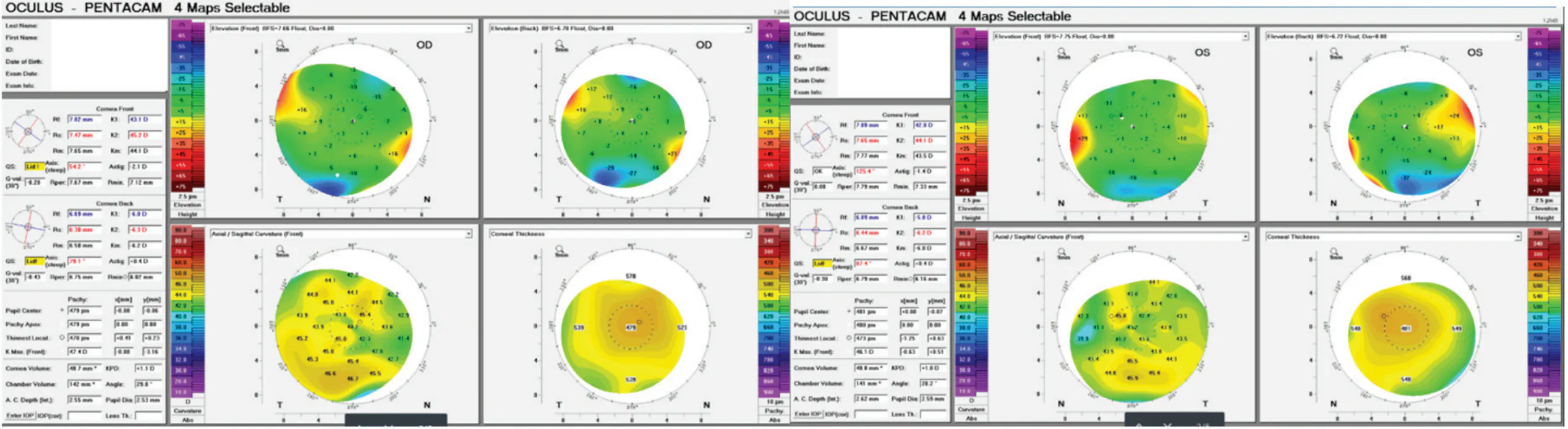
Figure 1. Corneal tomography (Pentacam, Oculus Optikgeräte) indicates a history of myopic LASIK and astigmatism.
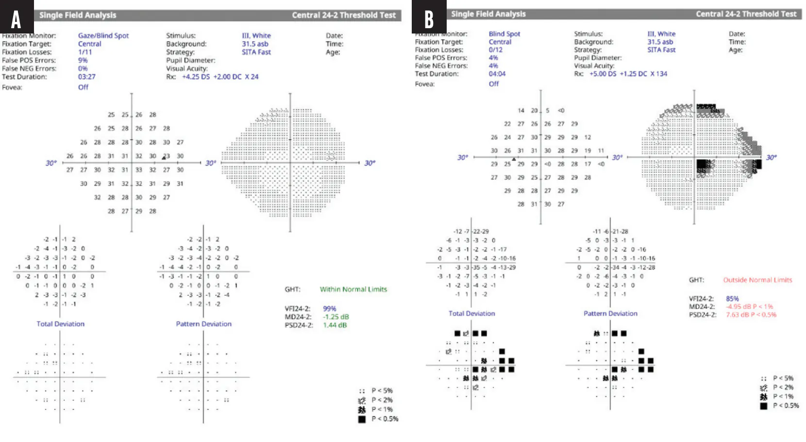
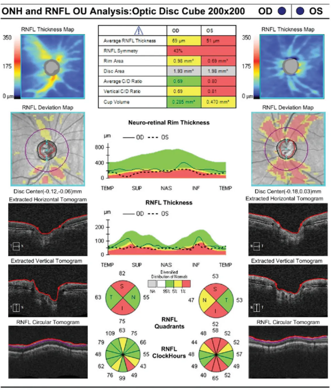
Visual field testing was essentially normal in the right eye but demonstrated paracentral, nasal, and superior glaucomatous defects in the left eye that corresponded to structural loss evident with OCT (Figures 2 and 3). The IOP was 12 mm Hg OD and 14 mm Hg OS. His current medical regimen for both eyes consisted of a fixed combination of dorzolamide and timolol administered twice daily and latanoprost instilled at night.
Selective laser trabeculoplasty was performed on both eyes, therapy with the fixed combination was halted, and aggressive lubrication of the ocular surface of each eye was initiated. Six weeks later, an improvement in the health of the ocular surface of each eye was observed, and the IOP measured 13 mm Hg OD and 15 mm Hg OS on latanoprost only.
IOL selection was then discussed. The goal was to balance the patient’s desire for spectacle independence with refractive predictability and postoperative visual quality. Given the asymmetric glaucomatous damage, epiretinal membrane, prior myopic LASIK, and astigmatism, a multifocal lens was not recommended. An extended depth of focus or enhanced monofocal IOL was a reasonable option. Given the paracentral glaucomatous loss in the left eye and previous refractive surgery, however, a monofocal Light Adjustable Lens (RxSight) was selected.
Cataract surgery was combined with a 360º canaloplasty and a 180º goniotomy using the Omni Edge (Sight Sciences). The postoperative Light Adjustable Lens adjustments targeted plano in the right eye and mild myopia (-0.50 D) in the left eye.
Two months after surgery, the patient’s UCVA was 20/20 OD and 20/30 OS at distance and J4 OU at near. He was able to use his phone without glasses and required over-the-counter readers only to read small print. Latanoprost was stopped postoperatively, and his IOP measured 10 mm Hg OU.
DISCUSSION
As this case demonstrates, individuals with glaucoma can have high visual expectations in addition to ocular comorbidities and a history of refractive surgery. Customizing refractive cataract surgery to the patient is essential. Optimizing the ocular surface preoperatively, reducing the medication burden, and educating patients on the benefits and limitations of advanced technology IOLs can help optimize their postoperative satisfaction.