Key Takeaways
- For patients with normal-tension glaucoma (NTG) whose disease continues to progress despite adequate IOP control, it is worth considering whether vascular risk factors are contributing to their optic nerve vulnerability.
- After confirming an NTG diagnosis, use a targeted vascular history to identify whether the patient may benefit from additional systemic evaluation, coordinate care with the patient's medical team, and continue to prioritize IOP reduction for NTG management.
- Evaluating vascular risk factors may improve understanding of disease progression and the overall health of patients with NTG.
A familiar clinical scenario in glaucoma practice is the patient with normal-tension glaucoma (NTG) whose disease continues to progress despite seemingly adequate IOP control. The instinct is often to pursue an even lower target IOP. Although IOP reduction remains the only intervention proven to slow disease progression,1 it is worth considering whether another factor could be contributing to the patient’s optic nerve vulnerability.
NTG accounts for a substantial proportion of primary open-angle glaucoma (POAG) cases worldwide, representing approximately 30% to 40% of POAG cases in Western populations and an even greater proportion in East Asian populations.2,3 The condition is characterized by progressive optic neuropathy despite untreated IOPs within a statistically normal range. Although IOP lowering remains the cornerstone of disease management, many patients continue to demonstrate structural or functional progression even after achieving their target pressures.4,5 This observation has prompted increasing interest in factors beyond IOP that may influence disease susceptibility and progression.
Among these, vascular dysfunction has received considerable attention. Proposed contributors include nocturnal hypotension, impaired vascular autoregulation, obstructive sleep apnea (OSA), vasospastic disorders, and other systemic conditions that may reduce optic nerve perfusion.6,7 Although these associations are supported by a growing body of observational evidence, it remains unclear whether identifying or treating these conditions alters the course of NTG.
This review outlines a practical approach to assessing vascular risk factors in patients with NTG, with a focus on the key clinical questions to ask and how the information acquired can be incorporated into disease evaluation and management.
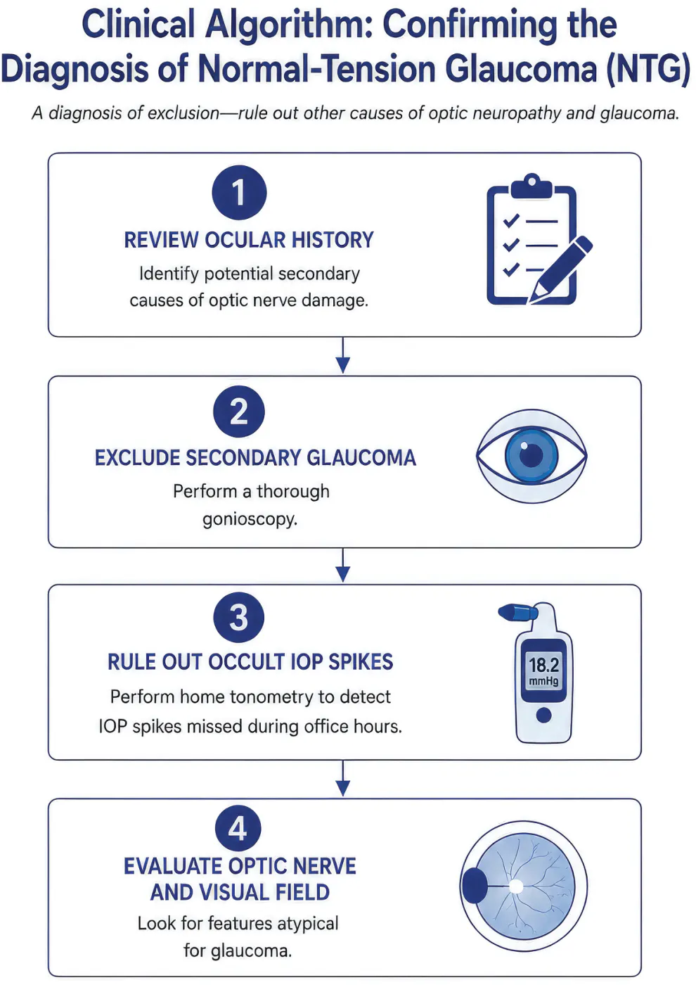
Figure 1. A practical algorithm for evaluating patients with NTG whose disease continues to progress despite apparently adequate IOP control.
IS IT TRULY NTG?
Before pursuing an extensive evaluation for vascular risk factors, it is worth confirming the diagnosis of NTG (Figure 1). Although NTG is common, it remains a diagnosis of exclusion.8 Patients with nonglaucomatous optic neuropathies or secondary causes of glaucoma may present with progressive optic nerve damage despite normal IOP, and these diagnoses require fundamentally different evaluation and management.
The initial step is to exclude secondary glaucomas. Careful gonioscopy should be performed to identify signs of pigment dispersion syndrome, pseudoexfoliation, angle recession, neovascularization, synechiae, or chronic angle closure.8 A thorough ocular history is equally important, with particular attention given to prior ocular trauma, corticosteroid exposure, uveitis, or intraocular surgery that might explain optic nerve damage or influence IOP measurements.
Office-based measurements may not capture the patient’s full IOP pattern. In patients whose glaucomatous progression seems disproportionate to consistently low clinic measurements, an office-based diurnal curve or short-term home tonometry trial may reveal early-morning or out-of-office IOP spikes.6,8 These findings may suggest that treatment should focus on controlling previously unrecognized pressure elevations rather than simply lowering the target IOP. If no meaningful spikes are identified and progression is confirmed, a lower target IOP may be necessary.
The optic nerve and visual field should then be assessed for features that are atypical for glaucoma. Optic disc pallor that exceeds the degree of cupping, reduced color vision or central visual acuity out of proportion to glaucomatous damage, an unexplained relative afferent pupillary defect, or visual field defects that respect the vertical rather than horizontal meridian should prompt consideration of an alternative diagnosis. Marked asymmetry between the eyes, particularly in the absence of corresponding differences in IOP, also warrants closer scrutiny.
Not every patient with NTG requires neuroimaging; however, MRI of the brain and orbits should be considered when the clinical picture is inconsistent with glaucoma, particularly in younger patients and those with rapid progression despite a substantial IOP reduction, unexplained vision loss, or associated neurologic symptoms.9 Confirming the diagnosis at the outset helps ensure that the subsequent evaluation for vascular risk factors is directed toward the appropriate patient population.
WHY LOOK BEYOND IOP?
Once the diagnosis of NTG has been confirmed, the next challenge is determining why some patients’ disease progresses even though their IOP is at target. The Collaborative Normal-Tension Glaucoma Study (CNTGS) demonstrated that a 30% IOP reduction significantly slowed visual field progression.4,5 Treatment should continue to focus on optimizing pressure control. However, if progression persists despite seemingly adequate IOP, factors beyond IOP alone may be influencing a patient’s susceptibility to optic nerve damage.
One proposed explanation is impaired ocular perfusion. Optic nerve health depends on a balance between IOP, systemic blood pressure (BP), and intact vascular autoregulation to maintain adequate blood flow.7 In susceptible individuals, fluctuations in systemic perfusion, recurrent nocturnal hypotension, or vascular dysregulation may reduce optic nerve perfusion and increase vulnerability to glaucomatous injury. Low diastolic perfusion pressure (< 50 mm Hg) has been associated with a higher prevalence of POAG. Nocturnal mean arterial pressure reductions of more than 10 mm Hg below daytime levels may predict NTG progression.7 Although these mechanisms are biologically plausible and supported by observational studies, causality remains unproven.7
For the practicing clinician, the value of identifying vascular risk factors is not necessarily to uncover a new glaucoma treatment but rather to better understand the individual patient and optimize their care through collaboration. A history of OSA, migraine, Raynaud phenomenon, or symptomatic hypotension may help to explain continued progression despite low IOP, identify systemic conditions that warrant medical attention, and inform conversations with the patient’s primary care physician (PCP) or other specialists. The following sections outline a focused approach to screening for these conditions during routine glaucoma visits (Figure 2).
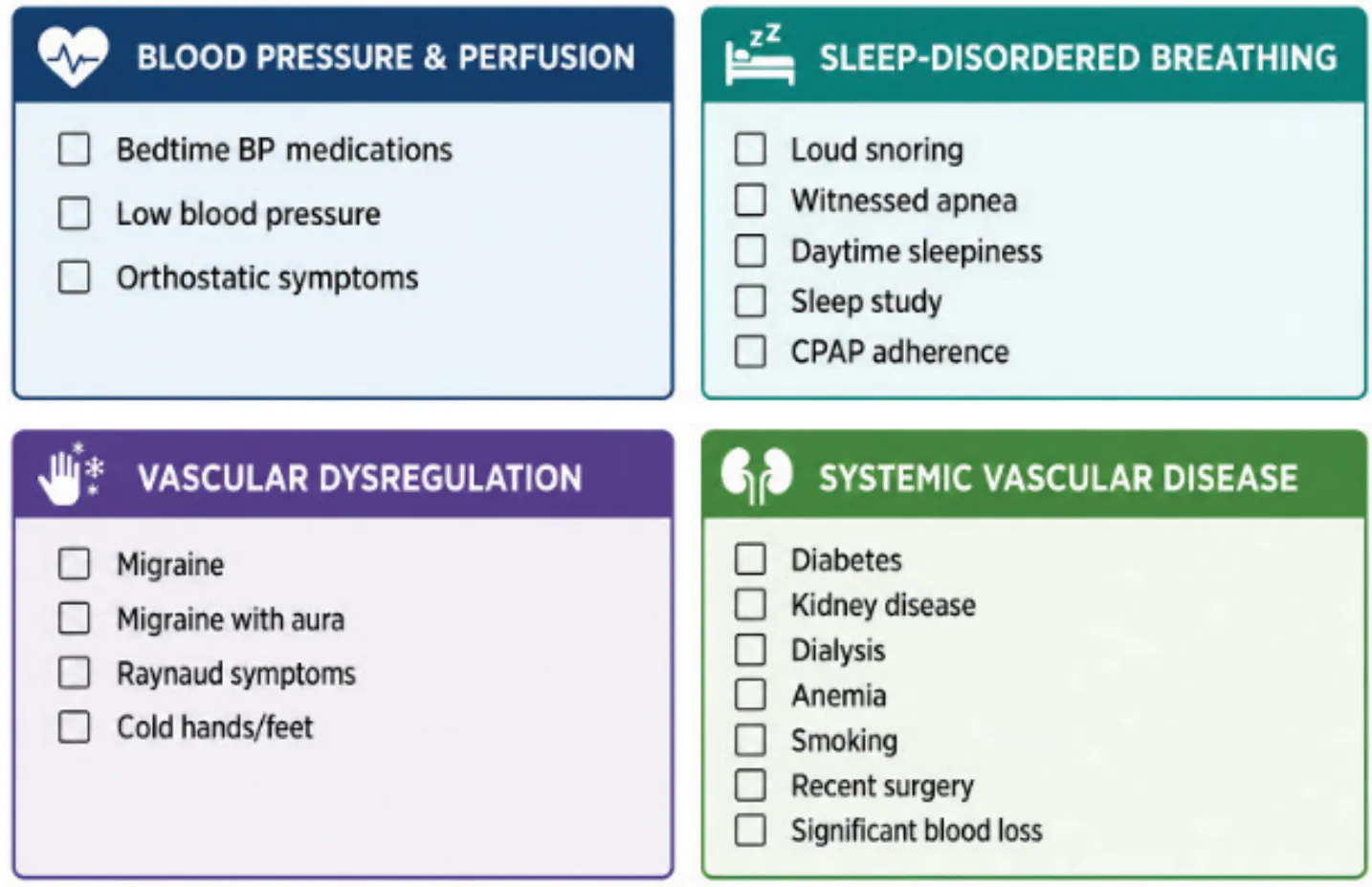
Figure 2. Targeted questions for identifying vascular and systemic risk factors in patients with NTG. Abbreviation: CPAP = continuous positive airway pressure.
A TARGETED VASCULAR HISTORY
BP AND NOCTURNAL HYPOTENSION
BP should be among the first considerations when evaluating a patient with progressive NTG. The goal is not simply to identify hypotension but to determine whether systemic perfusion may be compromised. Patients taking nighttime antihypertensive medications or reporting symptoms of hypotension may experience excessive reductions in nocturnal BP, potentially placing an already vulnerable optic nerve at greater risk of ischemia.6,7
Observational studies have consistently linked nocturnal hypotension with faster glaucomatous progression. In a systematic review and meta-analysis, excessive nocturnal systemic hypotension was associated with significantly greater odds of visual field progression, with odds ratios (ORs) of 3.32 (95% CI, 1.84–6.00) for systolic and 2.09 (95% CI, 1.20–3.64) for diastolic dipping.10 More recent studies have similarly found that patients with marked nocturnal BP reductions experienced more rapid central visual field loss than those with normal nocturnal BP patterns.11 Kwon et al demonstrated that nocturnal diastolic BP dips may be a more relevant predictor of visual field progression than systolic dips in patients with NTG.12 Although these studies did not establish causality, they support consideration of nocturnal hypotension as a potential contributor to progression.
Not every patient with NTG requires ambulatory BP monitoring. However, for patients whose disease continues to progress despite well-controlled IOP, particularly those reporting dizziness or nighttime antihypertensive use, it may be reasonable to discuss 24-hour BP monitoring with the patient’s PCP or cardiologist.10,13 Importantly, antihypertensive therapy should not be modified solely for glaucoma management without careful consideration of the patient’s overall cardiovascular health.
Questions to Ask the Patient:
- Do you take BP medications in the evening?
- Have you been told your BP runs low?
- Do you become lightheaded when standing?
Clinical Takeaways
Consider ambulatory BP monitoring only in patients with progressive NTG and symptoms suggestive of nocturnal hypotension. Any changes to antihypertensive therapy should be made in coordination with the patient’s PCP or cardiologist.
SLEEP-DISORDERED BREATHING
Sleep-disordered breathing, particularly OSA, is an important consideration in patients with progressive NTG. Recurrent episodes of nocturnal hypoxia, fluctuations in BP, and impaired vascular autoregulation have all been proposed as mechanisms by which OSA may increase optic nerve susceptibility.14 Although a causal relationship has not been established, multiple observational studies have demonstrated an association between OSA and glaucoma.
A recent systematic review and meta-analysis including more than 4.5 million participants found that OSA was associated with greater odds of glaucoma in the unadjusted analysis (OR, 3.66; 95% CI, 1.70–7.90). After adjustments for age and systemic comorbidities, the association was attenuated, with up to approximately 40% greater odds of glaucoma.15 Other studies have reported more conservative associations after adjustments for confounders.16,17 Although these findings support screening for OSA in appropriate patients, there is currently no evidence that OSA treatment slows the progression of NTG.14,15 Nevertheless, identifying OSA may improve a patient’s overall cardiovascular and metabolic health while providing additional context for continued disease progression despite well-controlled IOP.
Questions to Ask the Patient:
- Do you snore loudly or stop breathing during sleep?
- Do you wake up feeling unrefreshed or excessively sleepy?
- Have you been evaluated for OSA?
- If you have OSA, are you using your continuous positive airway pressure machine consistently?
Clinical Takeaways
OSA is a common and frequently underrecognized comorbidity of NTG. Although current evidence does not support OSA treatment as a strategy to slow glaucomatous progression, screening for symptoms and referring appropriate patients for a sleep evaluation may improve their overall health.
VASCULAR DYSREGULATION
Migraine and Raynaud phenomenon are among the most frequently reported systemic conditions associated with NTG and may serve as clinical clues to underlying vascular dysregulation.7,18 Although neither condition is specific to glaucoma, their presence may help identify patients whose optic nerves are more susceptible to ischemic injury. In a study of more than 2,000 patients, Gramer et al found that migraine was significantly more frequent in NTG (21.4%) than in POAG with elevated IOP (13.1%, P = .01).18 Observational studies have similarly reported higher rates of migraine and Raynaud phenomenon among patients with NTG than among those with higher-pressure POAG or in the general population, although the strength of these associations varies across studies.7,18 Together, these findings support the concept that vascular dysregulation may contribute to optic nerve vulnerability in a subset of patients.
The presence of migraine or Raynaud phenomenon should not alter glaucoma treatment in isolation, and there is no evidence that treating these conditions slows glaucomatous progression. However, recognition of these comorbidities may provide additional context when evaluating patients who continue to progress despite well-controlled IOP and may prompt a broader discussion of systemic vascular health.
Questions to Ask the Patient:
- Do you have a history of migraine headaches?
- Do your fingers or toes change color or become painful when cold?
- Are your hands or feet always cold?
- Have you been diagnosed with Raynaud phenomenon?
Clinical Takeaways
Patients with migraine, Raynaud phenomenon, cold hands or feet, and systemic hypotension often share features of what has been described as Flammer syndrome, a phenotype characterized by primary vascular dysregulation.19 Regardless of whether this represents a distinct clinical entity, these findings may point toward increased optic nerve susceptibility and reinforce the importance of considering vascular factors in patients with progressive NTG.
SYSTEMIC VASCULAR DISEASE
Systemic conditions that impair vascular health, reduce oxygen delivery, or compromise systemic perfusion may further increase optic nerve vulnerability in susceptible patients. Diabetes mellitus, anemia, chronic kidney disease, significant cardiovascular disease, and cigarette smoking may all contribute to reduced vascular reserve, although their individual relationships with NTG remain less well established than those of nocturnal hypotension or vascular dysregulation.
In a case-control study, patients with NTG had higher rates of diabetes (OR, 3.01), anemia (OR, 2.18), peripheral vascular disease (OR, 2.61), systemic hypotension (OR, 4.43), and Raynaud syndrome (OR, 3.09) compared with matched controls.20 Rather than implicating any single condition as the cause of NTG, these findings suggest that optic nerve vulnerability may be among the cumulative effects of systemic vascular disease. This broader perspective might help explain why some patients’ glaucoma continues to progress despite seemingly adequate IOP control.
For most patients, identifying these comorbidities will not change glaucoma treatment directly. Instead, it should prompt collaboration with the patient’s PCP or other specialists to optimize the patient’s systemic health while continuing to focus on IOP reduction.
Questions to Ask the Patient:
- Do you have diabetes or kidney disease?
- Have you been told you are anemic?
- Are you receiving dialysis?
- Have you ever smoked?
- Have you recently undergone major surgery or experienced significant blood loss?
Clinical Takeaways
Diabetes, anemia, and chronic kidney disease are best viewed as comorbid conditions that may contribute to optic nerve susceptibility rather than direct causes of NTG. Recognizing these systemic diseases encourages a more holistic approach to patient care while maintaining IOP reduction as the cornerstone of glaucoma management.
DISC HEMORRHAGES: A CLUE NOT TO IGNORE
Among the clinical findings in NTG, an optic disc hemorrhage is one of the strongest predictors of future structural and functional progression.21,22 A recent Cochrane systematic review identified a disc hemorrhage as a clinically important prognostic factor for glaucomatous progression (hazard ratio, 2.03; 95% CI, 1.55–2.67),21 and a meta-analysis found a pooled OR of 5.15 (95% CI, 3.72–7.14) for NTG progression in eyes with a baseline disc hemorrhage.22 Lee et al proposed that a disc hemorrhage should be reclassified from a risk factor to an indicator of ongoing glaucomatous progression.23 Furthermore, nocturnal BP dipping has been identified as a risk factor for a disc hemorrhage occurrence in NTG.24
The detection of a disc hemorrhage should prompt a careful reassessment of the patient’s disease rather than an automatic change in therapy. Confirm there is genuine progression, reassess the appropriateness of the target IOP, and determine the need for closer surveillance with OCT and visual field testing.
Clinical Takeaways
A disc hemorrhage is a marker of active glaucomatous disease and should prompt closer surveillance and a reassessment of the overall management plan.
PUTTING IT INTO PRACTICE
When a patient with NTG continues to experience progression despite IOP control, the next step is not simply to add another medication but to pause and reassess the broader clinical picture. Physicians should confirm that the diagnosis is correct, determine whether progression is genuine, and revisit the patient’s history for vascular risk factors that may provide additional context for their disease. If progression is confirmed, the target IOP should be reassessed. Laser or surgical intervention may be considered when further pressure reduction is required and cannot be achieved with topical medical therapy alone.
A targeted vascular history can be incorporated into routine clinic visits with only a few additional questions. Screening for nocturnal hypotension, sleep-disordered breathing, migraine, Raynaud phenomenon, and systemic vascular disease may help identify patients who would benefit from further evaluation by their PCP or an appropriate specialist. Although the treatment of these comorbidities has not been shown to alter the course of NTG, recognizing them may improve overall patient care and help explain why some patients continue to experience progression despite well-controlled IOP.
CONCLUSION
Ultimately, evaluating vascular risk factors should complement, not replace, established glaucoma management. IOP reduction remains the only proven disease-modifying therapy for NTG.1,4,5 A thoughtful history, careful examination, and multidisciplinary approach allow clinicians to better understand the whole patient while continuing to deliver evidence-based glaucoma care. n
1. Weinreb RN, Aung T, Medeiros FA. The pathophysiology and treatment of glaucoma: a review. JAMA. 2014;311(18):1901-1911. doi:10.1001/jama.2014.3192
2. Kim KE, Park KH. Update on the prevalence, etiology, diagnosis, and monitoring of normal-tension glaucoma. Asia Pac J Ophthalmol (Phila). 2016;5(1):23-31. doi:10.1097/APO.0000000000000177
3. Zhao J, Solano MM, Oldenburg CE, et al. Prevalence of normal-tension glaucoma in the Chinese population: a systematic review and meta-analysis. Am J Ophthalmol. 2019;199:101-110. doi:10.1016/j.ajo.2018.10.017
4. Collaborative Normal-Tension Glaucoma Study Group. The effectiveness of intraocular pressure reduction in the treatment of normal-tension glaucoma. Am J Ophthalmol. 1998;126(4):498-505. doi:10.1016/S0002-9394(98)00272-4
5. Anderson DR; Normal Tension Glaucoma Study. Collaborative Normal Tension Glaucoma Study. Curr Opin Ophthalmol. 2003;14(2):86-90. doi:10.1097/00055735-200304000-00006
6. Jayaram H, Kolko M, Friedman DS, Gazzard G. Glaucoma: now and beyond. Lancet. 2023;402(10414):1788-1801. doi:10.1016/S0140-6736(23)01289-8
7. Fan N, Wang P, Tang L, Liu X. Ocular blood flow and normal tension glaucoma. Biomed Res Int. 2015;2015:308505. doi:10.1155/2015/308505
8. Gedde SJ, Bowden EC, Challa P, et al. Primary open-angle glaucoma preferred practice pattern. Ophthalmology. 2026;133(4):P1-P103. doi:10.1016/j.ophtha.2025.12.029
9. Kosior-Jarecka E, Wróbel-Dudzińska D, Pietura R, et al. Results of neuroimaging in patients with atypical normal-tension glaucoma. Biomed Res Int. 2020;2020:9093206. doi:10.1155/2020/9093206
10. Bowe A, Grünig M, Schubert J, et al. Circadian variation in arterial blood pressure and glaucomatous optic neuropathy: a systematic review and meta-analysis. Am J Hypertens. 2015;28(9):1077-1082. doi:10.1093/ajh/hpv016
11. Park J, Song WK, Yoon J, Kim KE, Kook MS. Fast central visual field progression in patients with normal-tension glaucoma and nocturnal blood pressure dip. Am J Ophthalmol. 2026;281:233-248. doi:10.1016/j.ajo.2025.09.018
12. Kwon J, Jo YH, Jeong D, Shon K, Kook MS. Baseline systolic versus diastolic blood pressure dip and subsequent visual field progression in normal-tension glaucoma. Ophthalmology. 2019;126(7):967-979. doi:10.1016/j.ophtha.2019.03.001
13. Charlson ME, de Moraes CG, Link A, et al. Nocturnal systemic hypotension increases the risk of glaucoma progression. Ophthalmology. 2014;121(10):2004-2012. doi:10.1016/j.ophtha.2014.04.016
14. García-Sánchez A, Villalaín I, Asencio M, García J, García-Rio F. Sleep apnea and eye diseases: evidence of association and potential pathogenic mechanisms. J Clin Sleep Med. 2022;18(1):265-278. doi:10.5664/jcsm.9552
15. Cheong AJY, Wang SKX, Woon CY, et al. Obstructive sleep apnoea and glaucoma: a systematic review and meta-analysis. Eye (Lond). 2023;37(15):3065-3083. doi:10.1038/s41433-023-02471-6
16. Han X, Lee SS, Ingold N, et al. Associations of sleep apnoea with glaucoma and age-related macular degeneration: an analysis in the United Kingdom Biobank and the Canadian Longitudinal Study on Aging. BMC Med. 2021;19(1):104. doi:10.1186/s12916-021-01973-y
17. Mazurek W, Mazurek Ł, Rękas-Mazurek B, Rękas M. The pathophysiological association between obstructive sleep apnea and glaucoma: a current update. J Clin Med. 2026;15(13):5215. doi:10.3390/jcm15135215
18. Gramer G, Weber BH, Gramer E. Migraine and vasospasm in glaucoma: age-related evaluation of 2027 patients with glaucoma or ocular hypertension. Invest Ophthalmol Vis Sci. 2015;56(13):7999-8007. doi:10.1167/iovs.15-17274
19. Konieczka K, Flammer J. Treatment of glaucoma patients with Flammer syndrome. J Clin Med. 2021;10(18):4227. doi:10.3390/jcm10184227
20. Funk RO, Hodge DO, Kohli D, Roddy GW. Multiple systemic vascular risk factors are associated with low-tension glaucoma. J Glaucoma. 2022;31(1):15-22. doi:10.1097/IJG.0000000000001964
21. Piyasena MP, Daka Q, Qureshi R, et al. Prognostic factors associated with progression of open-angle glaucoma in adults. Cochrane Database Syst Rev. 2025;12:CD015436. doi:10.1002/14651858.CD015436.pub2
22. Tan CQL, Tan CJ, Tai BC, et al. Longitudinal association between disc hemorrhage and primary open-angle glaucoma progression: a systematic review and meta-analysis. Ophthalmic Epidemiol. 2026;33(3):310-319. doi:10.1080/09286586.2025.2568395
23. Lee EJ, Kee HJ, Han JC, Kee C. Evidence-based understanding of disc hemorrhage in glaucoma. Surv Ophthalmol. 2021;66(3):412-422. doi:10.1016/j.survophthal.2020.09.001
24. Kwon J, Lee J, Choi J, Jeong D, Kook MS. Association between nocturnal blood pressure dips and optic disc hemorrhage in patients with normal-tension glaucoma. Am J Ophthalmol. 2017;176:87-101. doi:10.1016/j.ajo.2017.01.002